. 2015 Nov 1;195(9):4378-86.
doi: 10.4049/jimmunol.1501702. Epub 2015 Sep 25.

Tissue Distribution of Memory T and B Cells in Rhesus Monkeys following Influenza A Infection

Affiliations

Tissue Distribution of Memory T and B Cells in Rhesus Monkeys following Influenza A Infection

Sathit Pichyangkul et al. J Immunol. .

Abstract

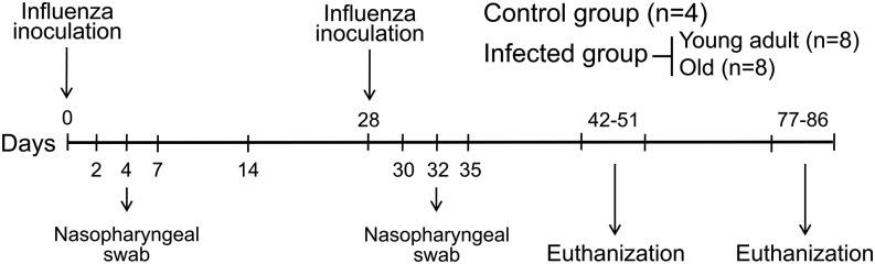
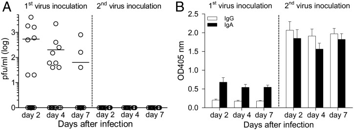
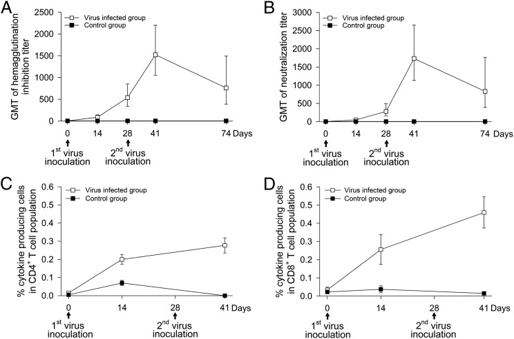
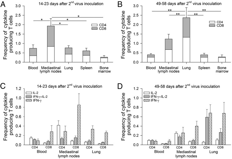
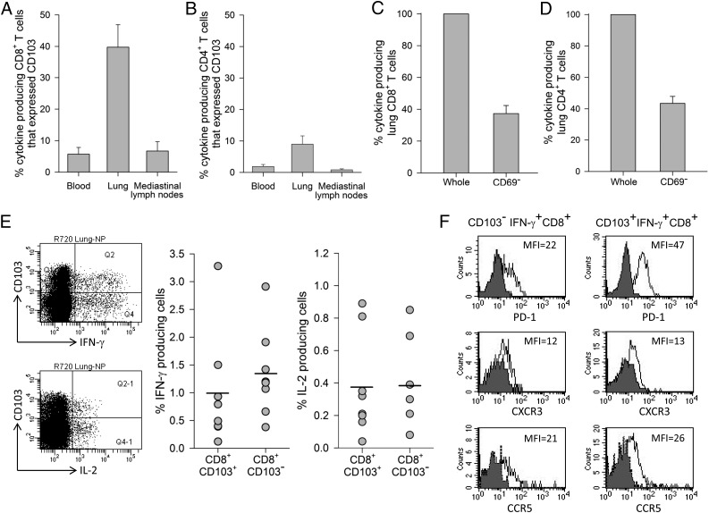
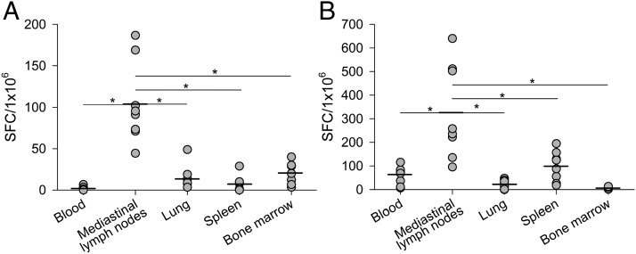
Studies of influenza-specific immune responses in humans have largely assessed systemic responses involving serum Ab and peripheral blood T cell responses. However, recent evidence indicates that tissue-resident memory T (TRM) cells play an important role in local murine intrapulmonary immunity. Rhesus monkeys were pulmonary exposed to 2009 pandemic H1N1 virus at days 0 and 28 and immune responses in different tissue compartments were measured. All animals were asymptomatic postinfection. Although only minimal memory immune responses were detected in peripheral blood, a high frequency of influenza nucleoprotein-specific memory T cells was detected in the lung at the "contraction phase," 49-58 d after second virus inoculation. A substantial proportion of lung nucleoprotein-specific memory CD8(+) T cells expressed CD103 and CD69, phenotypic markers of TRM cells. Lung CD103(+) and CD103(-) memory CD8(+) T cells expressed similar levels of IFN-γ and IL-2. Unlike memory T cells, spontaneous Ab secreting cells and memory B cells specific to influenza hemagglutinin were primarily observed in the mediastinal lymph nodes. Little difference in systemic and local immune responses against influenza was observed between young adult (6-8 y) and old animals (18-28 y). Using a nonhuman primate model, we revealed substantial induction of local T and B cell responses following 2009 pandemic H1N1 infection. Our study identified a subset of influenza-specific lung memory T cells characterized as TRM cells in rhesus monkeys. The rhesus monkey model may be useful to explore the role of TRM cells in local tissue protective immunity after rechallenge and vaccination.


Figures

FIGURE 1.
FIGURE 1.
Study design for influenza inoculation and tissue collection. Rhesus monkeys were exposed via intranasal and intratracheal inoculation to 2009 pH1N1 virus at days 0 and 28. Peripheral blood, mediastinal lymph nodes, lung, spleen, and bone marrow were collected at days 14–23 and 49–58 after second virus inoculation and assessed for immune responses.
FIGURE 2.
FIGURE 2.
Replication of 2009 pH1N1 in rhesus monkeys and the presence of HA-specific IgG and IgA in nasal secretions. (A) Virus replication in nasal secretions of 16 infected animals was measured by plaque assay. Each data point represents the value for an individual monkey and horizontal lines are means. (B) ELISA was used to measure HA-specific IgG and IgA in nasal secretions (1:4 dilution) and expressed as OD at 405 nm. Data shown are mean ± SE.
FIGURE 3.
FIGURE 3.
Kinetics of serum Ab and peripheral blood T cell responses. Ab responses were measured by HAI (A) and NT (B) assays and each data point represents the GMT ± SE. Peripheral blood CD4+ (C) and CD8+ (D) T cell responses were assessed by in vitro T cell recall assay against influenza NP. Intracellular cytokine staining was used to assess the frequencies of NP-specific cytokine-secreting T cells. Stained cells were analyzed by multicolor flow cytometry. Data shown are means ± SE of percent cytokine-producing cells (IFN-γ-, IL-2-, and IFN-γ plus IL-2) in CD4+ or CD8+ T cell population.
FIGURE 4.
FIGURE 4.
Generation of NP-specific memory CD4+ plus CD8+ T cells in different tissue compartments (peripheral blood, mediastinal lymph nodes, lung, spleen, and bone marrow) at the expansion phase (14–23 d after the second virus inoculation) (A) and the contraction phase (49–58 d after the second virus inoculation) (B). Data shown are means ± SE (*p < 0.05, **p < 0.01, Wilcoxon test). Cytokine profiles of NP-specific memory CD4+ and CD8+ T cells at the expansion phase (C) and contraction phase (D) were also evaluated by separating cells into three distinct populations based on the production of IL-2 alone, IFN-γ alone, and IFN-γ plus IL-2. Data shown are means ± SE.
FIGURE 5.
FIGURE 5.
Expression of CD103 and CD69, phenotypic markers of TRM on NP-specific lung memory T cells at the contraction phase. Percentage of cytokine producing CD8+ (A) or CD4+ (B) memory T cells that expressed CD103 in peripheral blood, lung, and mediastinal lymph nodes. Depletion of CD69-positive cells from lung mononuclear immune cells and cytokine response in NP-specific CD8+ (C) or CD4+ (D) memory T cells. Means ± SE of n = 8 are shown. Representative flow cytometry plot of lung CD103+ and CD103CD8+ T cells expressing IFN-γ and IL-2 and frequencies of IFN-γ and IL-2 production from lung CD103+ and CD103CD8+ T cells from eight animals (E). Each data point represents the value for an individual monkey and horizontal lines are means. Phenotype of IFN-γ–producing CD8+ T cells in the lung (F). Shaded histograms represent isotype control and solid lines represent lung CD103 or CD103+IFNγ+CD8+ T cells expressing PD-1, CXCR3, and CCR5. Data are representative of four monkeys.
FIGURE 6.
FIGURE 6.
Generation of HA-specific spontaneous ASCs and memory B cells at the contraction phase. Mononuclear immune cells isolated from different tissue compartments were assayed for the presence of spontaneous ASCs (A) and memory B cells (B) specific to influenza HA using ELISPOT assay. Each data point represents value for an individual monkey and horizontal lines are means (*p < 0.01, Wilcoxon test).

References

    1. Osterholm M. T., Kelley N. S., Sommer A., Belongia E. A. 2012. Efficacy and effectiveness of influenza vaccines: a systematic review and meta-analysis. Lancet Infect. Dis. 12: 36–44. - PubMed
    1. Hobson D., Curry R. L., Beare A. S., Ward-Gardner A. 1972. The role of serum haemagglutination-inhibiting antibody in protection against challenge infection with influenza A2 and B viruses. J. Hyg. (Lond.) 70: 767–777. - PMC - PubMed
    1. Black S., Nicolay U., Vesikari T., Knuf M., Del Giudice G., Della Cioppa G., Tsai T., Clemens R., Rappuoli R. 2011. Hemagglutination inhibition antibody titers as a correlate of protection for inactivated influenza vaccines in children. Pediatr. Infect. Dis. J. 30: 1081–1085. - PubMed
    1. Gravenstein S., Drinka P., Duthie E. H., Miller B. A., Brown C. S., Hensley M., Circo R., Langer E., Ershler W. B. 1994. Efficacy of an influenza hemagglutinin-diphtheria toxoid conjugate vaccine in elderly nursing home subjects during an influenza outbreak. J. Am. Geriatr. Soc. 42: 245–251. - PubMed
    1. Yap K. L., Ada G. L. 1978. Cytotoxic T cells in the lungs of mice infected with an influenza A virus. Scand. J. Immunol. 7: 73–80. - PubMed

Publication types

MeSH terms