Review
. 2015 Oct;41(7):682-90.
doi: 10.1055/s-0035-1564044. Epub 2015 Sep 26.

Structure-Function Relationship of the Interaction between Tissue Factor and Factor VIIa

Affiliations
Review

Structure-Function Relationship of the Interaction between Tissue Factor and Factor VIIa

Joshua M Gajsiewicz et al. Semin Thromb Hemost. 2015 Oct.

Abstract

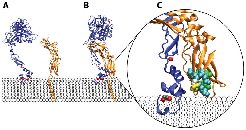
Interactions between tissue factor and factor VIIa are the primary initiators of coagulation in hemostasis and certain thrombotic diseases. Tissue factor, an integral membrane protein expressed extensively outside of the vasculature, is the regulatory protein cofactor for coagulation factor VIIa. Factor VIIa, a trypsin-like serine protease homologous with other blood coagulation proteases, is weakly active when free in solution and must bind its membrane-bound cofactor for physiologically relevant activity. Tissue factor allosterically activates factor VIIa by several mechanisms such as active site positioning, spatial stabilization, and direct interactions with the substrate. Protein-membrane interactions between tissue factor, factor VIIa, and substrates all play critical roles in modulating the activity of this enzyme complex. Additionally, divalent cations such as Ca(2+) and Mg(2+) are critical for correct protein folding, as well as protein-membrane and protein-protein interactions. The contributions of these factors toward tissue factor-factor VIIa activity are discussed in this review.


Figures

Figure 1
Figure 1
Crystal structures of FVIIa and sTF arranged on a membrane surface. A) FVIIa (blue) has an extended conformation and binds to anionic phospholipids in membrane bilayers through its N-terminal GLA-domain (depicted here in contact with the membrane). Coordination of divalent cations such as Ca2+ (red spheres) and Mg2+ (gray spheres) by the GLA domain is critical for proper domain folding and function. In addition, a Ca2+ ion is bound to the first EGF-like domain of FVIIa and also to the protease domain of this protein (the domain farthest from the membrane). The isolated ectodomain of TF (sTF, orange) is depicted here as anchored to the membrane surface via a single transmembrane helix, which has been modeled in. Full-length TF also contains a 21 amino acid-long cytoplasmic tail (not shown) which is implicated in interactions with the cytoskeleton. B) Crystal structure of the sTF-FVIIa complex, with the transmembrane helix of TF modeled in. FVIIa interacts extensively with sTF, with a binding interface that spans all domains of FVIIa and sTF. C) Close-up of TF residues putatively involved in substrate recognition (i.e., the substrate-binding exosite region of TF). In addition to allosterically activating FVIIa, TF is thought to directly interact with the protein substrates, FIX and FX, through membrane-proximal residues. Thus, TF residues Tyr157, Lys159, Ser163, Gly164, Lys165, Lys166, and Tyr185 (shown as van Der Waals radii and colored according to identity as follows; Teal: Lysine, White: Glycine, Yellow: Serine, Green: Tyrosine) contribute significantly to interactions with substrate as demonstrated by mutagenesis studies. (Panel C is rotated ~45° from panel B.) The structure of the sTF-FVIIa complex in panels B and C is rendered from pdb file 3TH2 using VMD Molecular Graphics Viewer. The isolated structures of FVIIa and sTF shown in panel A are a separation of the two from the TF-FVIIa complex. The transmembrane helix attached to sTF is adopted from pdb file 1A11.

References

    1. Mackman N. The role of tissue factor and factor VIIa in hemostasis. Anesth Analg. 2009;108(5):1447–1452. - PMC - PubMed
    1. Morrissey JH. Tissue factor: a key molecule in hemostatic and nonhemostatic systems. Int J Hematol. 2004;79(2):103–108. - PubMed
    1. Drake TA, Morrissey JH, Edgington TS. Selective cellular expression of tissue factor in human tissues. Implications for disorders of hemostasis and thrombosis. Am J Pathol. 1989;134(5):1087–1097. - PMC - PubMed
    1. Fleck RA, Rao LVM, Rapaport SI, Varki N. Localization of human tissue factor antigen by immunostaining with monospecific, polyclonal anti-human tissue factor antibody. Thromb Res. 1990;59(2):421–437. - PubMed
    1. Wilcox JN, Smith KM, Schwartz SM, Gordon D. Localization of tissue factor in the normal vessel wall and in the atherosclerotic plaque. Proc Natl Acad Sci U S A. 1989;86(8):2839–2843. - PMC - PubMed

Publication types