. 2015 Nov;5(11):1164-1177.
doi: 10.1158/2159-8290.CD-15-0369. Epub 2015 Sep 26.

Genomic Characterization of Brain Metastases Reveals Branched Evolution and Potential Therapeutic Targets

Affiliations

Genomic Characterization of Brain Metastases Reveals Branched Evolution and Potential Therapeutic Targets

Priscilla K Brastianos et al. Cancer Discov. 2015 Nov.

Abstract

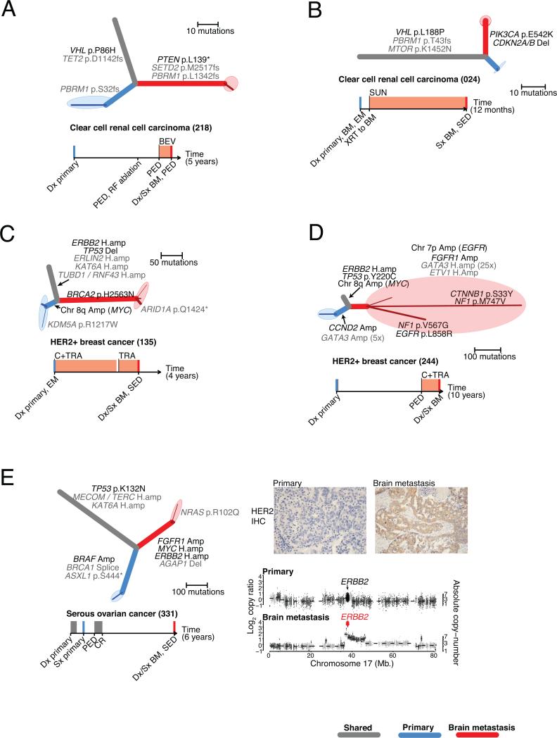
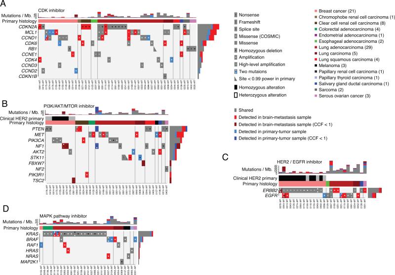
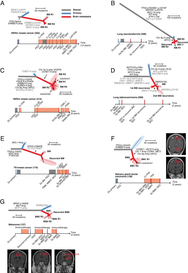
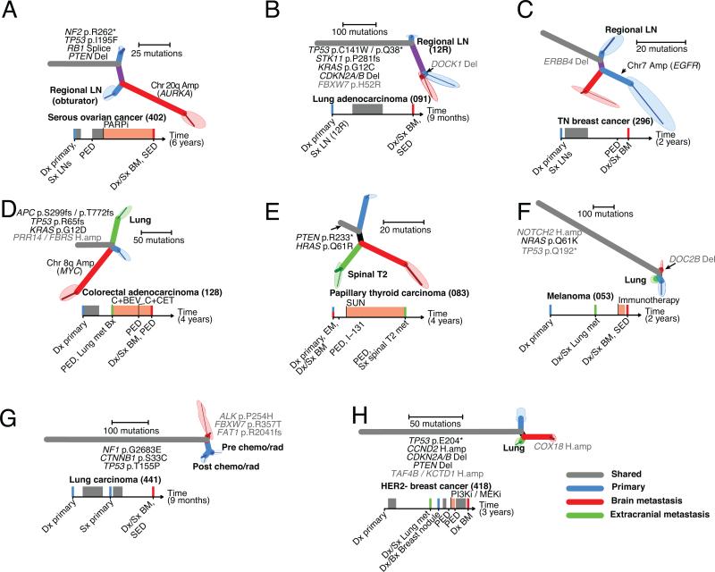
Brain metastases are associated with a dismal prognosis. Whether brain metastases harbor distinct genetic alterations beyond those observed in primary tumors is unknown. We performed whole-exome sequencing of 86 matched brain metastases, primary tumors, and normal tissue. In all clonally related cancer samples, we observed branched evolution, where all metastatic and primary sites shared a common ancestor yet continued to evolve independently. In 53% of cases, we found potentially clinically informative alterations in the brain metastases not detected in the matched primary-tumor sample. In contrast, spatially and temporally separated brain metastasis sites were genetically homogenous. Distal extracranial and regional lymph node metastases were highly divergent from brain metastases. We detected alterations associated with sensitivity to PI3K/AKT/mTOR, CDK, and HER2/EGFR inhibitors in the brain metastases. Genomic analysis of brain metastases provides an opportunity to identify potentially clinically informative alterations not detected in clinically sampled primary tumors, regional lymph nodes, or extracranial metastases.

Significance: Decisions for individualized therapies in patients with brain metastasis are often made from primary-tumor biopsies. We demonstrate that clinically actionable alterations present in brain metastases are frequently not detected in primary biopsies, suggesting that sequencing of primary biopsies alone may miss a substantial number of opportunities for targeted therapy.


Figures

Figure 1
Figure 1. Brain metastases harbor clinically actionable mutations not detected in primary-tumor samples
A-E. Phylogenetic trees inferred for five example cases. Branch colors indicate the types of tissue samples descended from each branch (grey: shared by all samples, blue: primary-tumor sample, red: brain metastasis). Darker-colored lines correspond to subpopulations of cancer cells detected with CCF < 1; the maximally branching evolutionary relationships of these clusters are drawn on the ends of each sample branch, surrounded by shaded ellipses denoting the tissue sample. The thickness of each branch is proportional to the CCF of mutations on that branch. Potentially clinically informative (TARGET) alterations (black) and additional likely oncogenic alterations (grey) are annotated onto the phylogenetic branches on which they occurred. Timelines depict the sequence of diagnosis, treatment, and tissue sampling for each case, with chemotherapy treatment intervals denoted by grey rectangles, and treatment with specified targeted agents denoted by orange rectangles. Colored vertical lines denote collection of sequenced cancer tissues (blue: primary, red: brain metastasis). BEV, bevacizumab; BM, brain metastasis; BM1, brain metastasis from one anatomic location; BM2, brain metastasis from second anatomic location; Bx, biopsy; C, chemotherapy; CET, cetuximab; CR, complete response; Dx, diagnosis; EM, extracranial metastasis; I-131, radioactive iodine; LAP, lapatinib; LN, lymph node; PARPi, PARP inhibitor; PBM, progressive brain metastasis; PED, progressive extracranial disease; PI3Ki, PI3K inhibitor; SED, stable extracranial disease; Sx, surgery; SUN, sunitinib; TRA, trastuzumab; WBRT, whole brain radiation therapy; XRT, radiation E. Also shows immunohistochemical staining (IHC) for HER2 in samples of the primary tumor (left), and brain metastasis (right). In addition, genomic copy-ratios on Chromosome 17 are shown (bottom) for the primary-tumor sample (top) and brain metastasis (bottom). Large diamonds correspond to exons of ERBB2, colored according to amplification status (black: unamplified, red: amplified).
Figure 2
Figure 2. The landscape of potentially clinically actionable alterations in brain metastases and primary-tumor samples
A-D. Alterations in genes (rows) that may predict sensitivity to the indicated class of targeted agent. Vertical columns correspond to cases, which are ordered by primary histology and presence/absence of alterations. Stacked bar-graphs indicating the number of somatic point-mutations detected in each phylogenetic branch of each case (columns) are shown at the top of each panel. HER2 status determined during clinical evaluation is denoted by black: positive, grey: negative, white: not measured.
Figure 3
Figure 3. Anatomically and regionally distinct brain metastasis samples share actionable drivers
A-G. Seven cases for which multiple regionally separated or anatomically distinct brain metastases were sequenced. The samples labeled R1, R2, etc. refer to different regions of the same pathology block. Phylogenetic trees and clinical histories are shown for each case as in Figure 2. C,F., minor subclones shared by > 1 tissue sample were detected (as described in the methods). For these cases, the shared areas denote the tissue samples, and indicate which subclones are present in each sample. F,G. Gadolinium-enhanced MRIs of the sampled brain metastases are shown.
Figure 4
Figure 4. Regional lymph nodes and distal extracranial metastases are not a reliable surrogate for actionable mutation in brain metastases
A-H. Eight cases for which at least one primary tumor sample, regional lymph node and extracranial metastasis were sequenced. Phylogenetic trees and clinical histories are shown for each case as in Figure 2. Tissue samples from extracranial metastases are depicted in green.

References

    1. Brastianos PK, Curry WT, Oh KS. Clinical discussion and review of the management of brain metastases. J Natl Compr Canc Netw. 2013;11:1153–64. - PubMed
    1. Eichler AF, Chung E, Kodack DP, Loeffler JS, Fukumura D, Jain RK. The biology of brain metastases-translation to new therapies. Nat Rev Clin Oncol. 2011;8:344–56. - PMC - PubMed
    1. Campbell PJ, Yachida S, Mudie LJ, Stephens PJ, Pleasance ED, Stebbings LA, et al. The patterns and dynamics of genomic instability in metastatic pancreatic cancer. Nature. 2010;467:1109–13. - PMC - PubMed
    1. Gerlinger M, Rowan AJ, Horswell S, Larkin J, Endesfelder D, Gronroos E, et al. Intratumor heterogeneity and branched evolution revealed by multiregion sequencing. N Engl J Med. 2012;366:883–92. - PMC - PubMed
    1. Liu W, Laitinen S, Khan S, Vihinen M, Kowalski J, Yu G, et al. Copy number analysis indicates monoclonal origin of lethal metastatic prostate cancer. Nat Med. 2009;15:559–65. - PMC - PubMed

Publication types

MeSH terms