

Development of Retinal Layers in Prenatal Human Retina
- PMID: 26410132
- PMCID: PMC5538359
- DOI: 10.1016/j.ajo.2015.09.023
Development of Retinal Layers in Prenatal Human Retina
Abstract
Purpose: To determine the developmental sequence of retinal layers to provide information on where in utero pathologic events might affect retinal development.
Design: Qualitative and quantitative descriptive research.
Methods: A histology collection of human eyes from fetal week (Fwk) 8 to postnatal (P) 10 weeks was analyzed. The length of the nasal and temporal retina was measured along the horizontal meridian in 20 eyes. The location of the inner plexiform layer (IPL) and outer plexiform layer (OPL) was identified at each age, and its length measured.
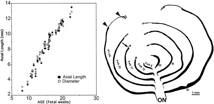
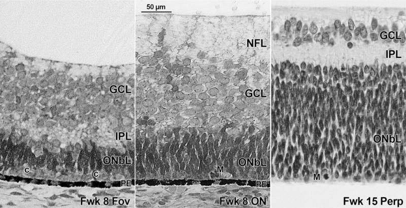
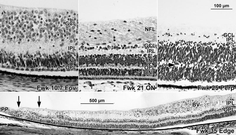
Results: The human eye retinal length increased from 5.19 mm at Fwk 8 to 20.92 mm at midgestation to 32.88 mm just after birth. The IPL appeared in the presumptive fovea at Fwk 8, reached the eccentricity of the optic nerve by Fwk 12, and was present to both nasal and temporal peripheral edges by Fwk 18-21. By contrast, the OPL developed slowly. A short OPL was first present in the Fwk 11 fovea and did not reach the eccentricity of the optic nerve until midgestation. The OPL reached the retinal edges by Fwk 30. Laminar development of both IPL and OPL occurred before vascular formation.
Conclusions: In human fetal retina, the IPL reached the far peripheral edge of the retina by midgestation and the OPL by late gestation. Only very early in utero events could affect IPL lamination in the central retina, but events occurring after Fwk 20 in the peripheral retina would overlap OPL laminar development in outer retina.
Copyright © 2016 Elsevier Inc. All rights reserved.
Figures
References
-
- Mann I. The Development of the Human Eye. 3rd. New York: Grune & Stratton; 1964.
-
- Yuodelis C, Hendrickson A. A qualitative and quantitative analysis of the human fovea during development. Vision Res. 1986;26(6):847–855. - PubMed
-
- Xiao M, Hendrickson A. Spatial and temporal expression of short, long/medium or both opsins in human fetal cones. J Comp Neurol. 2000;425:545–559. - PubMed
Publication types
MeSH terms
Grants and funding
LinkOut - more resources
Full Text Sources
Other Literature Sources
Medical
