Review
. 2016 Apr;11(4):505-19.
doi: 10.1007/s11548-015-1292-0. Epub 2015 Sep 26.

CustusX: an open-source research platform for image-guided therapy

Affiliations
Review

CustusX: an open-source research platform for image-guided therapy

Christian Askeland et al. Int J Comput Assist Radiol Surg. 2016 Apr.

Abstract

Purpose: CustusX is an image-guided therapy (IGT) research platform dedicated to intraoperative navigation and ultrasound imaging. In this paper, we present CustusX as a robust, accurate, and extensible platform with full access to data and algorithms and show examples of application in technological and clinical IGT research.

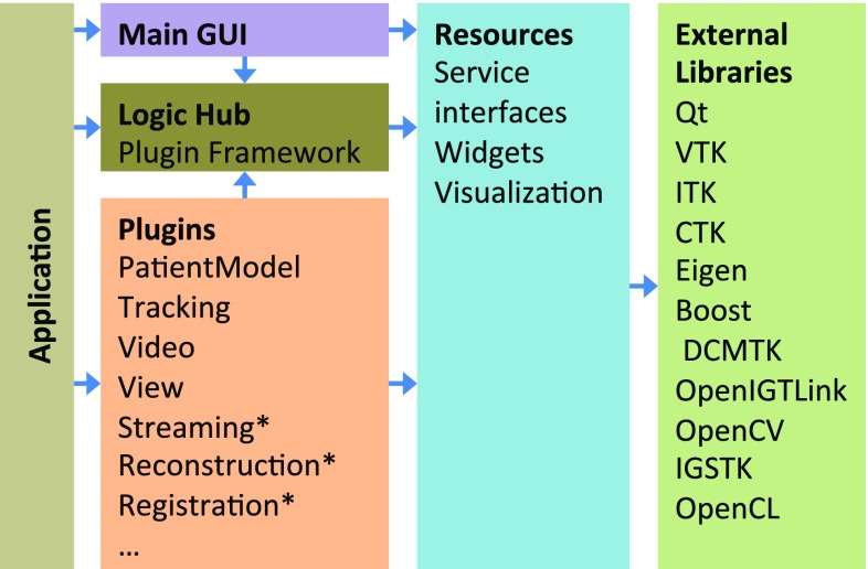
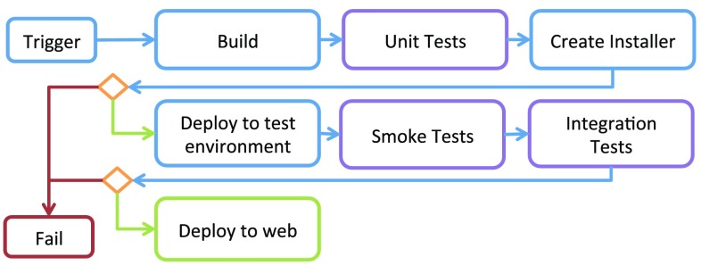
Methods: CustusX has been developed continuously for more than 15 years based on requirements from clinical and technological researchers within the framework of a well-defined software quality process. The platform was designed as a layered architecture with plugins based on the CTK/OSGi framework, a superbuild that manages dependencies and features supporting the IGT workflow. We describe the use of the system in several different clinical settings and characterize major aspects of the system such as accuracy, frame rate, and latency.

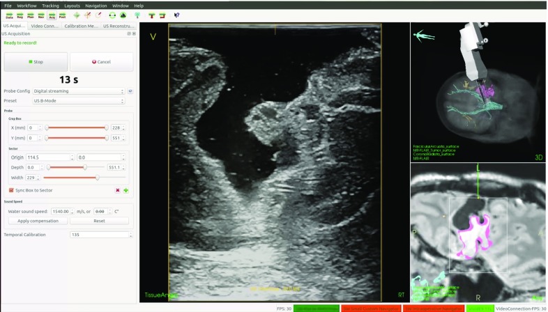
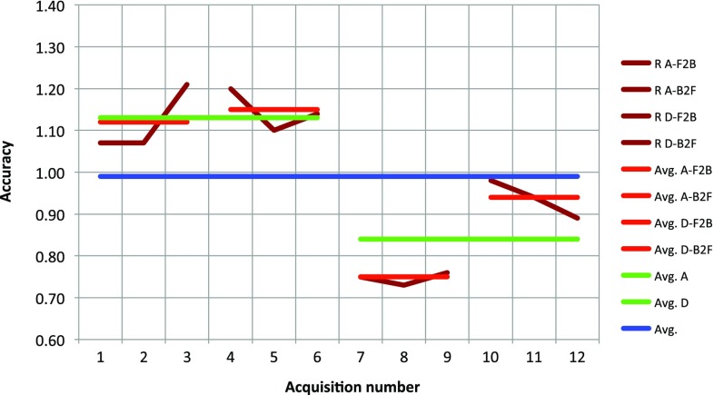
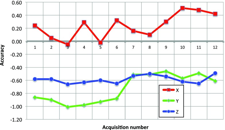
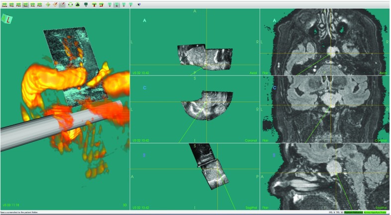
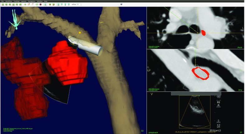
Results: The validation experiments show a navigation system accuracy of [Formula: see text]1.1 mm, a frame rate of 20 fps, and latency of 285 ms for a typical setup. The current platform is extensible, user-friendly and has a streamlined architecture and quality process. CustusX has successfully been used for IGT research in neurosurgery, laparoscopic surgery, vascular surgery, and bronchoscopy.

Conclusions: CustusX is now a mature research platform for intraoperative navigation and ultrasound imaging and is ready for use by the IGT research community. CustusX is open-source and freely available at http://www.custusx.org.

Keywords: Computer-assisted interventions; Image-guided therapy; Intraoperative ultrasound; Open source; Platform.


Figures

Fig. 1
Fig. 1
Main window. The virtual patient is shown in the central view area, using a combination of 2D and 3D views. Other user interaction components such as widgets and toolbars are also available
Fig. 2
Fig. 2
CustusX architecture. Arrows show the dependency direction. The application initialize the plugins and the user interface, with the Logic Hub as a plugin repository. Plugins contribute to functionality and extensibility in a modular way. The plugins marked with an asterisk have multiple instances. Resources and components from external libraries are used as building blocks throughout the system
Fig. 3
Fig. 3
A normalized clinical workflow as used by CustusX. The steps model a procedure from data preparation and registration through intraoperative navigation and image acquisition, ending in postoperative analysis
Fig. 4
Fig. 4
An example setup of CustusX for use in neurosurgery, with a tracked US probe imaging the patient
Fig. 5
Fig. 5
Ultrasound acquisition during brain tumor surgery
Fig. 6
Fig. 6
Continuous integration flow. The flow is triggered by either a commit, a nightly timer, or a manual request. The system is then built and tested in both the build folder and after deployment. Failure at any stage aborts the flow. If all steps succeed, the binaries are published on a Web server
Fig. 7
Fig. 7
Navigation system accuracy (NSA) based on preoperative (p) MR (red line) and intraoperative (i) US (green line). iUS can be used to correct pMR using various image-to-image registration techniques (blue line)
Fig. 8
Fig. 8
Automatic method for evaluating the accuracy in US-based navigation. a The phantom with a single wire cross in the middle of the water tank and a reference frame in the front, and a US probe imaging the wire. b 1-mm-diameter model of the nominal wire cross in green, and an US volume of the wire cross in red. A mismatch of about 1 mm can be seen c Centerlines of the nominal (green) and US (red) wire crosses. d ICP registration between the two centerlines, initial correspondence shown. e After convergence. US centerlines now match the nominal. f The displacement is equal to the NSA
Fig. 9
Fig. 9
Accuracy as a function of acquisitions performed under various conditions; both individual measurements and various averages are shown. See Table 1 for an explanation of the different labels used, R = NSA
Fig. 10
Fig. 10
Accuracy components in the reference frame of the ultrasound scan plane as a function of acquisition number. The directions are elevation (x), azimuth (y), and radial (z)
Fig. 11
Fig. 11
Intraoperative navigation using MR FLAIR and 3D US during resection of a low-grade glioma
Fig. 12
Fig. 12
Navigation and intraoperative 3D US in surgery of a pituitary tumor. Left column model of the distal part of the prototype US probe with the real-time 2D US image plane shown at its correct location, and the volume rendering of a 3D US power Doppler volume. Middle column orthogonal image slices reformatted from a 3D US image volume. Right column corresponding orthogonal image slices reformatted from a preoperative MR volume
Fig. 13
Fig. 13
CustusX 3D view during endovascular navigation showing cone beam CT image volume and real-time position tracking of catheter (red cylinder mark) and guidewire (yellow cylinder mark)
Fig. 14
Fig. 14
A navigation scene during EBUS-TBNA. Left 3D representation of the segmented airways, EBUS scope, lymph node, and tumor. Right Axial view of CT (top), view of CT corresponding to the US sector (middle), and US (bottom)

References

    1. Fedorov A, Beichel R, Kalpathy-Cramer J, Finet J, Fillion-Robin JC, Pujol S, Bauer C, Jennings D, Fennessy F, Sonka M, Buatti J, Aylward S, Miller JV, Pieper S, Kikinis R. 3D slicer as an image computing platform for the quantitative imaging network. Magn Reson Imaging. 2012;30(9):1323–1341. doi: 10.1016/j.mri.2012.05.001. - DOI - PMC - PubMed
    1. Lasso A, Heffter T, Rankin A, Pinter C, Ungi T, Fichtinger G. PLUS: open-source toolkit for ultrasound-guided intervention systems. IEEE Trans Biomed Eng. 2014;61(10):2527–2537. doi: 10.1109/TBME.2014.2322864. - DOI - PMC - PubMed
    1. Nolden M, Zelzer S, Seitel A, Wald D, Muller M, Franz AM, Maleike D, Fangerau M, Baumhauer M, Maier-Hein L, Maier-Hein KH, Meinzer HP, Wolf I. The medical imaging interaction toolkit: challenges and advances: 10 years of open-source development. Int J Comput Assist Radiol Surg. 2013;8(4):607–620. doi: 10.1007/s11548-013-0840-8. - DOI - PubMed
    1. Mercier L, Del Maestro RF, Petrecca K, Kochanowska A, Drouin S, Yan CX, Janke AL, Chen SJ, Collins DL. New prototype neuronavigation system based on preoperative imaging and intraoperative freehand ultrasound: system description and validation. Int J Comput Assist Radiol Surg. 2011;6(4):507–522. doi: 10.1007/s11548-010-0535-3. - DOI - PubMed
    1. Ince DC, Hatton L, Graham-Cumming J. The case for open computer programs. Nature. 2012;482(7386):485–488. doi: 10.1038/nature10836. - DOI - PubMed

Publication types

LinkOut - more resources