. 2016 Jun 2;35(22):2881-92.
doi: 10.1038/onc.2015.353. Epub 2015 Sep 28.

Estrogen promotes the brain metastatic colonization of triple negative breast cancer cells via an astrocyte-mediated paracrine mechanism

Affiliations

Estrogen promotes the brain metastatic colonization of triple negative breast cancer cells via an astrocyte-mediated paracrine mechanism

C A Sartorius et al. Oncogene. .

Abstract

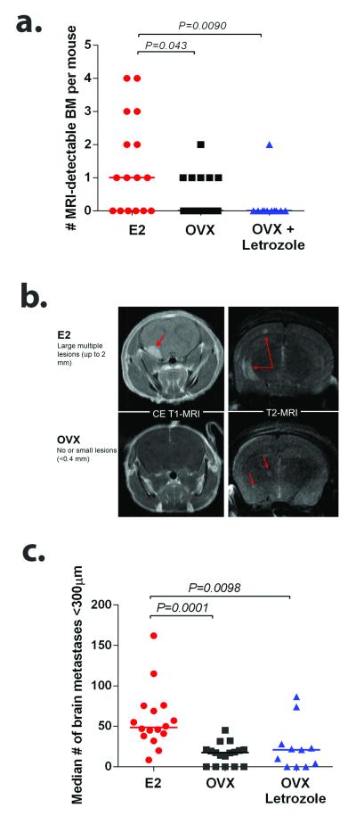
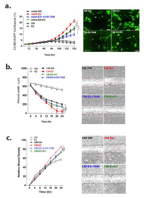
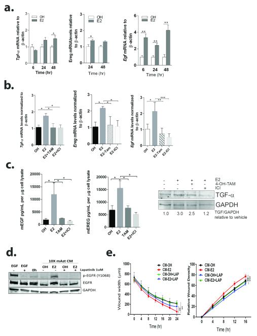
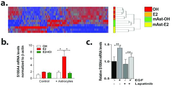
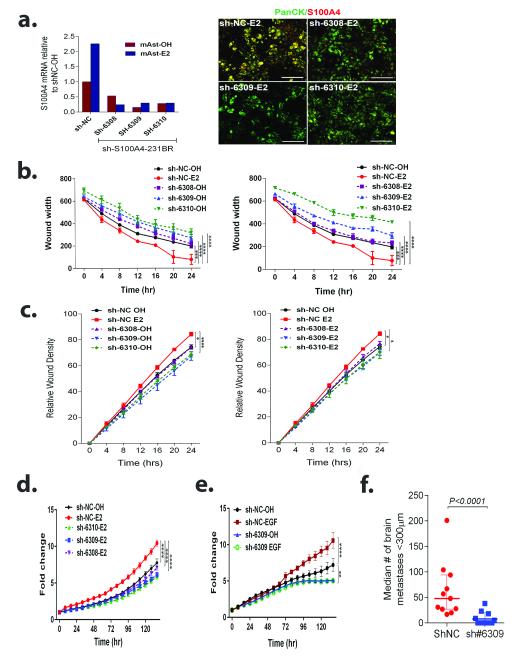
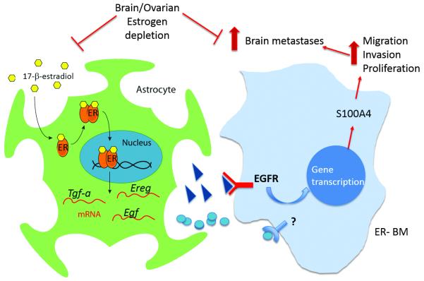
Brain metastases (BM) are a devastating consequence of breast cancer. BM occur more frequently in patients with estrogen receptor-negative (ER-) breast cancer subtypes; HER2 overexpressing (HER2+) tumors and triple-negative (TN) (ER-, progesterone receptor-negative (PR-) and normal HER2) tumors. Young age is an independent risk factor for the development of BM, thus we speculated that higher circulating estrogens in young, pre-menopausal women could exert paracrine effects through the highly estrogen-responsive brain microenvironment. Using a TN experimental metastases model, we demonstrate that ovariectomy decreased the frequency of magnetic resonance imaging-detectable lesions by 56% as compared with estrogen supplementation, and that the combination of ovariectomy and letrozole further reduced the frequency of large lesions to 14.4% of the estrogen control. Human BM expressed 4.2-48.4% ER+ stromal area, particularly ER+ astrocytes. In vitro, E2-treated astrocytes increased proliferation, migration and invasion of 231BR-EGFP cells in an ER-dependent manner. E2 upregulated epidermal growth factor receptor (EGFR) ligands Egf, Ereg and Tgfa mRNA and protein levels in astrocytes, and activated EGFR in brain metastatic cells. Co-culture of 231BR-EGFP cells with E2-treated astrocytes led to the upregulation of the metastatic mediator S100 Calcium-binding protein A4 (S100A4) (1.78-fold, P<0.05). Exogenous EGF increased S100A4 mRNA levels in 231BR-EGFP cells (1.40±0.02-fold, P<0.01 compared with vehicle control) and an EGFR/HER2 inhibitor blocked this effect, suggesting that S100A4 is a downstream effector of EGFR activation. Short hairpin RNA-mediated S100A4 silencing in 231BR-EGFP cells decreased their migration and invasion in response to E2-CM, abolished their increased proliferation in co-cultures with E2-treated astrocytes and decreased brain metastatic colonization. Thus, S100A4 is one effector of the paracrine action of E2 in brain metastatic cells. These studies provide a novel mechanism by which estrogens, acting through ER+ astrocytes in the brain microenvironment, can promote BM of TN breast cancers, and suggests existing endocrine agents may provide some clinical benefit towards reducing and managing BM.


Figures

Figure 1
Figure 1. Estrogen depletion decreases experimental breast cancer BM formation
OVX nude mice supplemented with placebo (OVX, n=17), estrogen (E2,n=16) or letrozole (OVX+Letrozole, n=11) were injected intracardially with 175,000 triple-negative 231BR-EGFP cells. Data represents two separate experiments. a. Number of MRI-detectable BM per mice in each group. Line designates group median. b. Representative contrast-enhanced (CE) T1-weighted and fat-suppressed T2-weighted MRI showing increased number and size of BM in E2-treated mice compared to OVX mice. c. Number of micrometastases per brain section (<300 μm). Each dot represents the median per mouse and the line designates the group median. Data was analyzed using non-parametric One Way ANOVA followed by Dunn’s multiple comparisons test. Graph shows adjusted P values.
Figure 2
Figure 2. Astrocytes in the brain metastatic microenvironment are ER+
a. ERα is detected in stroma (S) surrounding tumor cells (T) in clinical immunostains from human BM from ER+ and ER− subtypes. Graph shows the percent ERα positive-stained stromal area in immunostained human breast cancer BM (BCBM) (n=12), analyzed using the Aperio system (see Supplementary Figure 1). Colored dots represent different subtypes. b. Activated astrocytes (GFAP+) surrounding human BM express ERα and ERβ (green). ERβ is expressed in astrocytes and tumor cells in ER-case. c. GFAP+ mouse astrocytes express ERα and ERβ (green) in experimental BM of 231BR-EGFP cells. d. Left: Western blot shows expression of ERs in mouse primary astrocytes (mAst), ER+ MCF-7 cells, ER− MDA-MB-231 cells and ER− brain metastatic derivatives 231BR-EGFP and 231BR-HER2. α-tubulin is used as loading control. Right: Primary mouse astrocytes are reactive in vitro (GFAP+, red) and express ERα and ERβ (green). DAPI stains nuclei (blue).
Figure 3
Figure 3. E2-treated astrocytes increase proliferation, migration and invasion of brain metastatic cells in an ER-dependent manner
a. 231BR-EGFP cells were plated on top of a mouse-primary astrocyte monolayer and cultured in serum-free media with vehicle (OH) or E2 alone (10 nM) or E2 in combination with 1 μM 4-OH-Tam (E2+TAM) or 100 nM ICI (E2+ICI). 231BR-EGFP cells were also treated with vehicle (OH) or 10 nM E2 (E2) in 5% CS-FBS media in absence of astrocytes to determine direct effects of E2. Graph shows the average percentage of 231BR-EGFP density ± SEM (n=4 per treatment), representative of at least two independent experiments. P<0.01 CM-OH vs CM-E2 start at 120 h. Right: Representative image shows 231BR-EGFP cells at 6 days. b. 231BR-EGFP cells were serum-starved overnight and 10X CM of astrocytes treated for 72 h with vehicle (CM-OH) or 10 nM E2 alone (CM-E2) or in combination with 1 μM Tam (CM E2+TAM) or 100 nM ICI (CM E2+ICI), were used as chemoattractant in scratch wound assays. Graph shows wound width (μm) over time. CM-OH vs CM-E2 P<0.01 for each time point after 16 h. Right: Representative image of wound at 24 h. c. 231BR-EGFP cells were treated as in b, in a modified scratch wound assays (see methods). Graphs shows average relative wound confluence (RWC) ± SEM. RWC is proportional to the amount of cells invading through the ECM-filled wound. CM-OH vs CM-E2 P<0.05 start at 16hr. Right: Representative image of wound at 24hrs. For all graphs, **P<0.01, ***P<0.001, ****P<0.0001 indicates the significance at the latest time point in repeated measures ANOVA followed by post-hoc multiple comparisons test.
Figure 4
Figure 4. E2 upregulates EGFR ligands in astrocytes and results in EGFR activation in brain metastatic cells
a. Primary mouse astrocytes were treated with vehicle (OH) or 10 nM E2 for the indicated times and qRT-PCR was used to measure Egf, Tgfα and Ereg mRNA levels. Bars represent fold change mRNA levels normalized to mouse β-Actin mRNA and relative to OH-treated. *P<0.05, **P<0.01 vs. OH-control. n=3. b. Astrocytes were cultured in serum-free media containing vehicle (OH) or E2 alone (10 nM) or E2 in combination with 1 μM 4-OH-Tam (E2+TAM) or 100 nM ICI (E2+ICI) for qRT-PCR at 24 h (for Egf and Ereg) or 48 h (for Tgfα). Graph shows mRNA levels normalized to mouse β-actin relative to OH-treated astrocytes. Bars are average ± SEM. c. Astrocytes were cultured as in b for 48 h, and cell lysates assessed by ELISA (mEGF, mEREG) or Western blot (TGFα). Graphs shows mEGF and mEREG concentration in 1 μg total cell lysate. Bars are average ± SEM. WB shows precursor TGFα (16KD), GAPDH was used as loading control. Numbers indicate fold change of TGFα/GAPDH relative to OH-treated cells. d. 231BR-EGFP cells were starved for 12 h, treated with 1 uM lapatinib or vehicle (DMSO) for 6 h and then stimulated with 10 ng/ml EGF, CM-OH or CM-E2 for 10 min. GAPDH was used as loading control. e. 231BR-EGFP cells were plated in uncoated (left, migration) or matrigel-coated (right, invasion) plates, serum-starved for 16 h and treated with 1 μM lapatinib for 3 h. After scratch wound, invasion well were coated with matrigel and then CM-OH or CM-E2 used as chemoattractant. Graphs show average wound width ±SEM (migration) and average RWD ± SEM (invasion). For all graphs, *P<0.05, **P<0.01, ***P<0.001.
Figure 5
Figure 5. Paracrine action of estrogen through astrocytes results in upregulation of metastasis mediator S100A4 in 231BR-EGFP cells
a. Hierarchical clustering of genes significantly affected by E2-treated astrocytes in sorted 231BR-EGFP cells treated with OH, 10 nM E2 alone, or in co-culture with OH-treated (mASt-OH) or 10 nM E2-treated (mAst) mouse astrocytes. n=3 per group, P<0.05, 1.5 fold change cutoff. b. 231BR-EGFP cells were treated with OH, 10 nM E2 alone or in combination with 100 nM ICI , either alone or in co-culture with astrocytes for 24 h. Sorted GFP+ cells were used for qRT-PCR analyses. Graph shows average S100A4 mRNA levels normalized to β-actin and relative to OH-treated cells ± SEM (n=3). Data representative of two independent experiments. c. 231BR-EGFP cells were starved for 16 h, pre-treated with 1 uM lapatinib or vehicle for 6 h and then treated with vehicle or 10 ng/ml EGF in 5% charcoal stripped-FBS containing media for 24 h. Graphs shows average S100A4 mRNA levels normalized to GAPDH and relative to vehicle-treated cells ± SEM, **P<0.01.
Figure 6
Figure 6. S100A4 knockdown blocks proliferation, migration and invasion of 231BR-EGFP cells in response to E2-treated astrocytes and inhibits brain colonization in vivo
a. Left: 231BR-EGFP cells expressing scramble control (sh-NC) or S100A4 targeting shRNAS (sh6308, sh6309, sh6310) were co-cultured with OH or E2-treated astrocytes for 24 h and then sorted. Graph shows S100A4 mRNA expression normalized to β-actin and relative to shNC-OH-treated control. Right: Cells were co-cultured as in a, and S100A4 expression assessed by IF. Panel shows merged images of S100A4 (red) and pan-cytokeratin labeling 231BR-EGFP cells (green). Individual channels are shown in Supplementary figure 3. b. shNC or shS100A4 cells were serum-starved overnight and CM-OH or CM-E2 was used as chemoattractant in scratch wound assays. Graph shows average wound width ± SEM. c. shNC or shS100A4 cells were treated as in b, in a modified scratch wound assay. Graphs shows relative average wound confluence (RWC) ± SEM. d. shNC or shS100A4 cells were plated on top of an astrocyte monolayer and cultured in serum-free media with OH, E2, or E2+ICI, and GFP+ confluence measured over time using live imaging. Data represents fold change in GFP+cells confluence relative to day 0± SEM. e. shNC or shS100A4 cells were plated in 5% CSFBS containing media and treated with vehicle or 10 ng/mL EGF for 6 days. Graph shows fold change in GFP+ cells relative to time 0 ± SEM (n=4). For all graphs, **P<0.01, ***P<0.001, ****P<0.0001 indicates the significance at the latest time point in repeated measures ANOVA followed by post-hoc multiple comparisons test. f. Female OVX nude mice supplemented with estrogen pellets, were injected intracardially with 175,000 sh-NC (n=11) or shS100A4-6309 (n=12) 231BR-EGFP (Sh#6309) cells and mice euthanized 28 days after injection. Micrometastases (<300 μm) per brain section in 6 sections through the right hemisphere were quantified. Each dot represents the median per mouse and the line designates the group median. Data was analyzed using Mann-Whitney non-parametric t-test.
Figure 7
Figure 7. A simplified scheme of the paracrine effects of estrogen in brain metastases
Consistent with the notion that breast cancer cells benefit from normal homeostasis mechanisms at the metastatic site (48, 50), our data supports a novel mechanism whereby brain metastatic cells exploit estrogen signaling though ER+ astrocytes. Estrogen can activate ERs on astrocytes, leading to transcriptional upregulation of EGFR ligands, activation of EGFR on TN EGFR+ brain metastatic breast cancer cells, and upregulation of several metastatic mediators, including S100A4. Given the complex bi-directional interactions between astrocytes and breast cancer cells, it is possible that other factors regulated by ERs in astrocytes and receptors on brain metastatic cells participate in this process. Ovarian and local estrogen depletion decrease brain metastatic colonization of TN EGFR+ 231BR cells in vivo, emphasizing the need to consider the brain microenvironment in designing novel therapies for brain metastases and the possibility to extend the use of aromatase inhibitors in the prevention of brain metastases in TN EGFR+ patients.

References

    1. Cheng X, Hung MC. Breast cancer brain metastases. Cancer metastasis reviews. 2007;26(3-4):635–43. - PubMed
    1. Brufsky AM, Mayer M, Rugo HS, Kaufman PA, Tan-Chiu E, Tripathy D, et al. Central nervous system metastases in patients with HER2-positive metastatic breast cancer: incidence, treatment, and survival in patients from registHER. Clinical cancer research : an official journal of the American Association for Cancer Research. 2011;17(14):4834–43. - PubMed
    1. Kodack DP, Askoxylakis V, Ferraro GB, Fukumura D, Jain RK. Emerging strategies for treating brain metastases from breast cancer. Cancer Cell. 2015;27(2):163–75. - PMC - PubMed
    1. Boogerd W, Vos VW, Hart AA, Baris G. Brain metastases in breast cancer; natural history, prognostic factors and outcome. J Neurooncol. 1993;15(2):165–74. - PubMed
    1. Braccini AL, Azria D, Thezenas S, Romieu G, Ferrero JM, Jacot W. Prognostic factors of brain metastases from breast cancer: Impact of targeted therapies. The Breast. 2013;22(5):993–8. - PubMed