. 2015 Oct 13;13(2):337-49.
doi: 10.1016/j.celrep.2015.08.075. Epub 2015 Sep 24.

Chromatin Architecture of the Pitx2 Locus Requires CTCF- and Pitx2-Dependent Asymmetry that Mirrors Embryonic Gut Laterality

Affiliations

Chromatin Architecture of the Pitx2 Locus Requires CTCF- and Pitx2-Dependent Asymmetry that Mirrors Embryonic Gut Laterality

Ian C Welsh et al. Cell Rep. .

Abstract

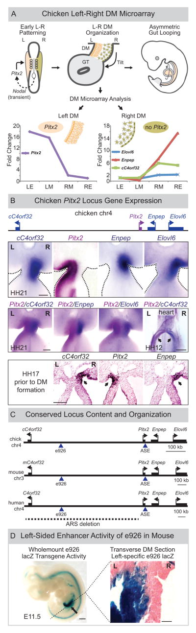
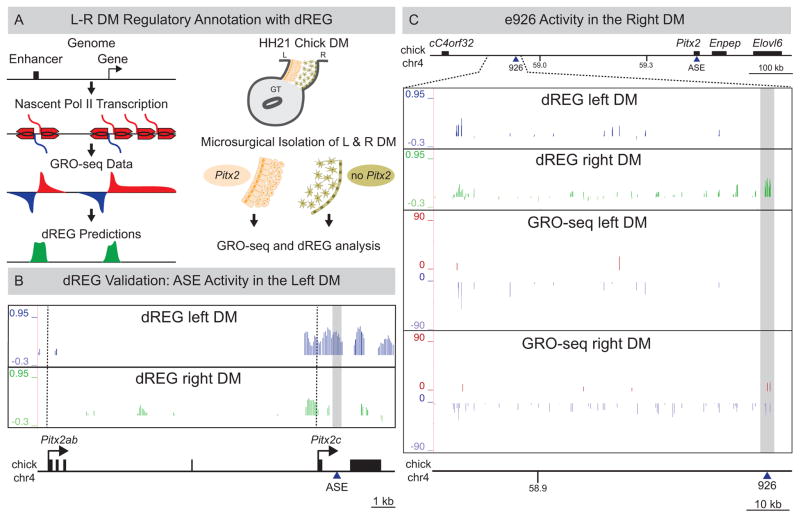
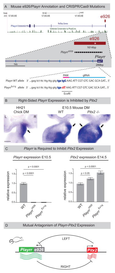
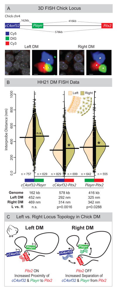
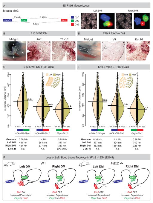
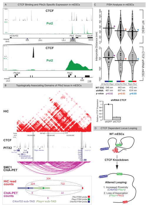
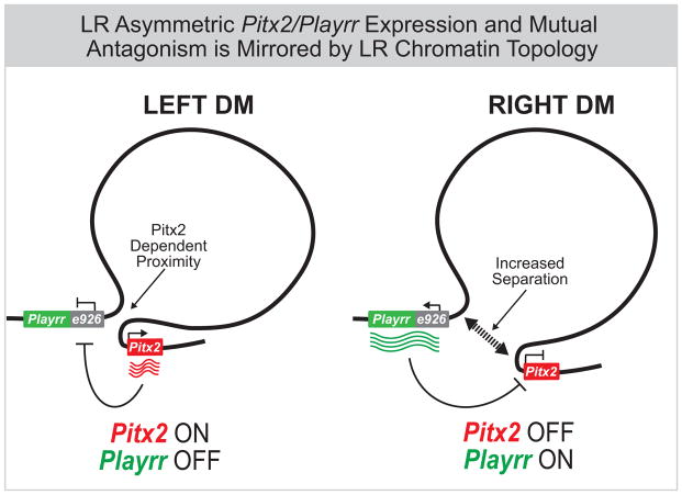
Expression of Pitx2 on the left side of the embryo patterns left-right (LR) organs including the dorsal mesentery (DM), whose asymmetric cell behavior directs gut looping. Despite the importance of organ laterality, chromatin-level regulation of Pitx2 remains undefined. Here, we show that genes immediately neighboring Pitx2 in chicken and mouse, including a long noncoding RNA (Pitx2 locus-asymmetric regulated RNA or Playrr), are expressed on the right side and repressed by Pitx2. CRISPR/Cas9 genome editing of Playrr, 3D fluorescent in situ hybridization (FISH), and variations of chromatin conformation capture (3C) demonstrate that mutual antagonism between Pitx2 and Playrr is coordinated by asymmetric chromatin interactions dependent on Pitx2 and CTCF. We demonstrate that transcriptional and morphological asymmetries driving gut looping are mirrored by chromatin architectural asymmetries at the Pitx2 locus. We propose a model whereby Pitx2 auto-regulation directs chromatin topology to coordinate LR transcription of this locus essential for LR organogenesis.


Figures

Figure 1
Figure 1. LR asymmetric gene expression at the Pitx2 locus
(A) Pitx2 initiates gut looping directed by LR changes in the DM cellular architecture (tan, left; green, right). DM microarray (LE, left epithelium; LM, left mesenchyme; RM, right mesenchyme; RE, right epithelium) identifies genes linked to Pitx2 with right side-restricted expression, validated in (B) via whole mount ISH (double labeling Pitx2, magenta; right-specific genes, blue) at HH21 (DM), at HH17 (lateral mesoderm, DM precursor, arrows) and at HH12 (sinus venosus of the primitive heart, arrows). (C) Pitx2 locus conservation in chicken, mouse, and human (dashed line, human ARS deletion). Note, human locus is inverted relative to its orientation on chr4. (D) e926 directs left-specific reporter gene expression (LacZ) in the left DM of transgenic mouse embryos. Scale bars: (B) 100μm, HH21 and HH17, 500μm, HH12; (D) 500μm, whole mount, 25μm, section. See also Figure S1.
Figure 2
Figure 2. LR DM GRO-seq/dREG analyses
(A) Left: GRO-seq and dREG. Right: Libraries were prepared from HH21 LR DM samples (B) dREG peaks corresponding to ASE are left-specific (grey box). Dashed vertical lines mark Pitx2ab and Pitx2c TSS. (C) dREG peaks overlapping e926 are right-specific (grey box) in contrast to left-specific e926 transgenic reporter activity. GRO-seq detects asymmetric transcription from the minus strand at the proximal gene desert in the right DM. See also Figure S2 and S3.
Figure 3
Figure 3. Identification of a conserved lncRNA, Playrr, asymmetrically transcribed from e926
(A) Annotation of mouse lncRNA Playrr (D030025E07Rik). e926 contains the TSS and exon 1 of Playrr and a conserved Pitx2 binding motif (asterisk). CRISPR/Cas9 mutation of e926/Playrr (PlayrrΔ926, 1420bp dashed line; PlayrrEx1sj, red and blue letters). Exon 1 (upper case), intron 1 (lower case), relative guide RNA (gRNA, light blue) and PAM motif (pink) were used to produce PlayrrEx1sj. (B) Whole mount ISH showing right-specific Playrr in WT HH21 chicken and E10.5 mouse embryos vs. bilateral Playrr expression in Pitx2-null mouse DM. (C) qRT-PCR expression in PlayrrΔ926 and PlayrrEx1sj mutants (n=3) relative to WT (n=3) of Playrr (left) and Pitx2 (right) at E10.5 or E14.5, respectively, error bars ± S.E.M. (D) Mutual antagonism of Playrr and Pitx2 expression. Scale bars in (B) 100μm. See also Figure S1 and S4.
Figure 4
Figure 4. Asymmetric chromatin interactions of the Pitx2 locus in the chicken LR DM
(A) Linear genomic distances in the chicken separating cC4orf32, Playrr, and Pitx2, marked with Cy5, DIG, and Cy3 labeled DNA probes, respectively. FISH labeled nuclei and individual loci in the left and right DM. (B) Split bean plots of interprobe distances for each probe pair (WT, HH21). Individual measurements are plotted as short horizontal ticks, the density trace and mean are plotted as the filled curve and large horizontal bar, respectively. Table summary of distances separating each probe pair in the linear genome or nucleus. (C) Model of Pitx2 locus organization and gene expression in the chicken DM. Scale bars: (A) 2μm and 300nm for nuclei and individual loci, respectively. See also Figure S4.
Figure 5
Figure 5. Spatial proximity of Playrr-Pitx2 chromatin interactions are dependent on Pitx2
(A) Linear genomic distances in the mouse separating mC4orf32, Playrr, and Pitx2, marked with Cy5, DIG, and Cy3 labeled DNA probes, respectively. FISH labeled nuclei and individual loci in the left and right DM. (B) Chiral looping of the WT midgut is accompanied by left-specific Isl1 and right-specific Tbx18. A, anterior; P, posterior; D, dorsal; V, ventral. (C) Split bean plots (WT E10.5). Table summary of distances separating each probe pair in the linear genome or nucleus. (D) Altered midgut looping and right-isomerized DM in Pitx2-null embryos is evidenced by loss of left-specific Isl1 and bilateral Tbx18 expression. (E) Split bean plots (Pitx2-null E10.5). (F) Model of Pitx2 locus organization and gene expression in the mouse DM. Scale bars: (A) 2μm and 300nm for nuclei and individual loci, respectively; (B) 250μm, whole mount, 25μm, section. See also Figure S4.
Figure 6
Figure 6. Playrr-Pitx2 chromatin interactions in mouse ES cells are dependent on CTCF
(A) Top: CTCF binding at the Pitx2 locus (CTCF ChIP-seq); Bottom: mESCs preferentially express the asymmetric Pitx2c isoform (RNA Pol II ChIP-seq). (B) A topologically associating domain (TAD) spans the Pitx2 locus (HiC, red; Smc1 ChIA-PET, magenta). Vertical grey bars highlight boundaries between sub-TADs. CTCF binding sites (ChIP-seq) at both Pitx2 and Playrr are associated with Pitx2 binding (Pitx2-FLAG ChIP-seq, asterisks). Quantification of HiC (red) and Smc1 ChIA-PET (magenta) interactions between the genomic interval containing the Pitx2 FISH probe and the Playrr sub-TAD (light green) compared to the mC4orf32 sub-TAD (light blue). (C) FISH analysis of Pitx2 locus topology in WT (top) or shRNA mediated CTCF knockdown mESCs (bottom, y-axis has been inverted); table shows summary of distances separating each probe pair in the nucleus. CTCF expression via qRT-PCR in control vs CTCF k.d. mESCs, error bars ± S.E.M. (D) Summary of Pitx2 locus topology in mESCs. See also Figure S7.
Figure 7
Figure 7. Summary of Pitx2 locus asymmetric expression and chromatin topology
In chicken and mouse, left-specific Pitx2 expression and right-sided expression of flanking genes at the Pitx2 locus, including the conserved lncRNA Playrr is accompanied by Pitx2 dependent asymmetric chromatin topology in the DM. In vivo, Pitx2 and Playrr expression are mutually antagonistic.

References

    1. Anderson E, Devenney PS, Hill RE, Lettice LA. Mapping the Shh long-range regulatory domain. Development. 2014 Oct;141(20):3934–3943. - PMC - PubMed
    1. Attanasio C, Nord AS, Zhu Y, Blow MJ, Li Z, Liberton DK, Morrison H, Plajzer-Frick I, Holt A, Hosseini R, et al. Fine tuning of craniofacial morphology by distant-acting enhancers. Science. 2013;342(6157):1241006. - PMC - PubMed
    1. Biswas D, Milne TA, Basrur V, Kim J, Elenitoba-Johnson KSJ, Allis CD, Roeder RG. Function of leukemogenic mixed lineage leukemia 1 (MLL) fusion proteins through distinct partner protein complexes. Proc Natl Acad Sci. 2011;108:15751–15756. - PMC - PubMed
    1. Bitoun E, Oliver PL, Davies KE. The mixed-lineage leukemia fusion partner AF4 stimulates RNA polymerase II transcriptional elongation and mediates coordinated chromatin remodeling. Hum Mol Genet. 2007;16:92–106. - PubMed
    1. Bonn S, Zinzen RP, Girardot C, Gustafson EH, Perez-Gonzalez A, Delhomme N, Ghavi-Helm Y, Wilczyński B, Riddell A, Furlong EEM. Tissue-specific analysis of chromatin state identifies temporal signatures of enhancer activity during embryonic development. Nat Genet. 2012;44:148–156. - PubMed

Publication types

Associated data