. 2015 Sep 28:6:8403.
doi: 10.1038/ncomms9403.

Structure of the native Sec61 protein-conducting channel

Affiliations

Structure of the native Sec61 protein-conducting channel

Stefan Pfeffer et al. Nat Commun. .

Abstract

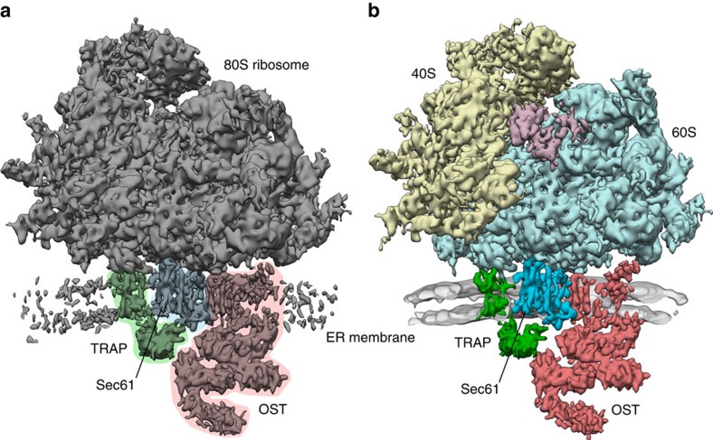
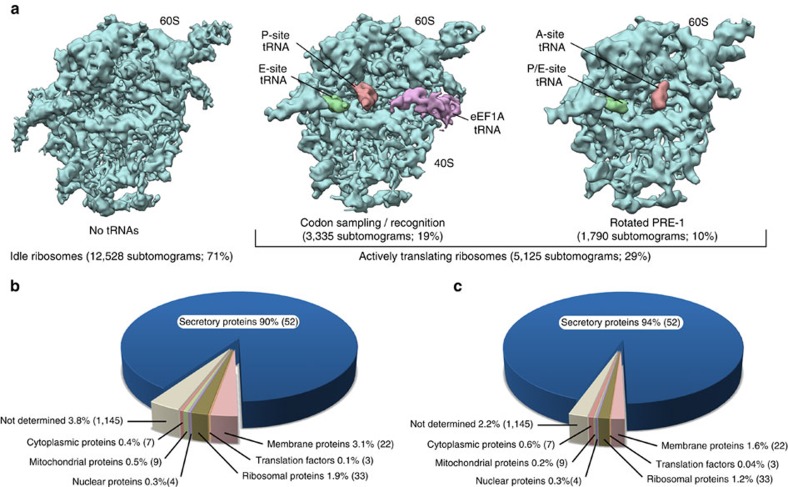
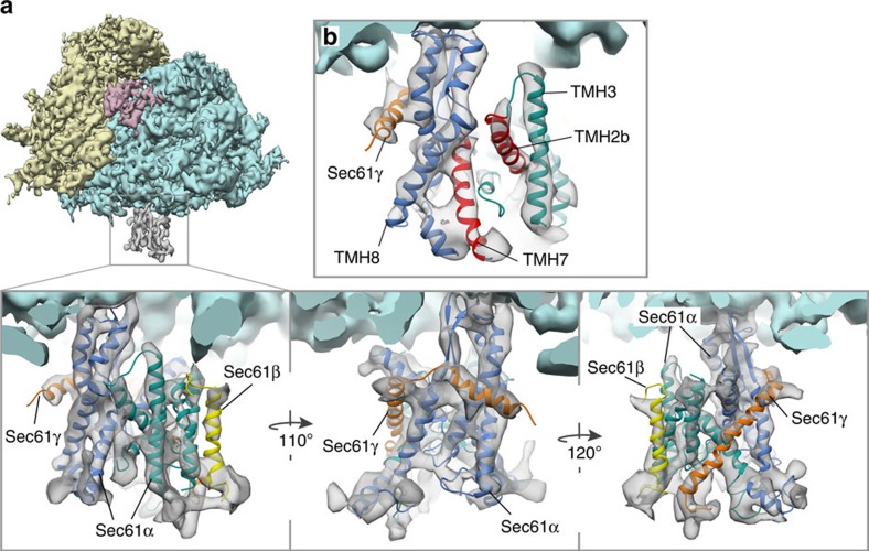
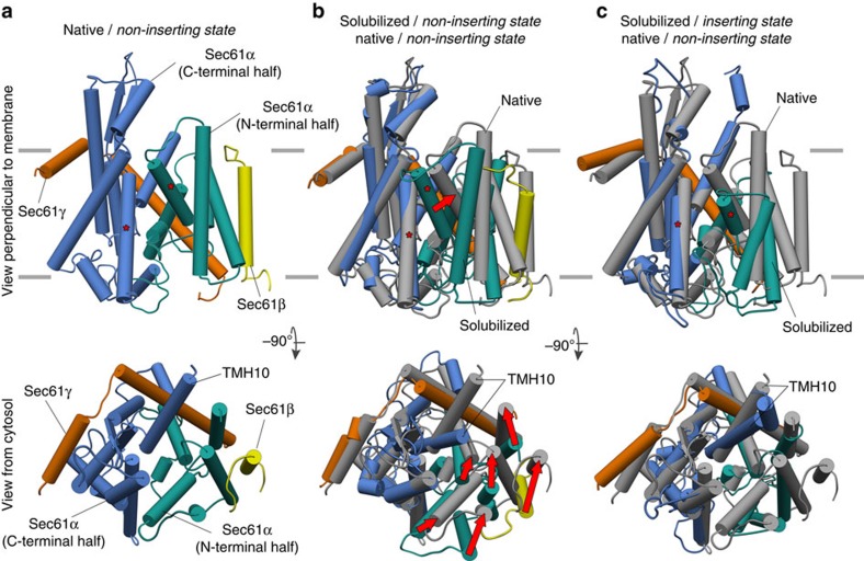
In mammalian cells, secretory and membrane proteins are translocated across or inserted into the endoplasmic reticulum (ER) membrane by the universally conserved protein-conducting channel Sec61, which has been structurally studied in isolated, detergent-solubilized states. Here we structurally and functionally characterize native, non-solubilized ribosome-Sec61 complexes on rough ER vesicles using cryo-electron tomography and ribosome profiling. Surprisingly, the 9-Å resolution subtomogram average reveals Sec61 in a laterally open conformation, even though the channel is not in the process of inserting membrane proteins into the lipid bilayer. In contrast to recent mechanistic models for polypeptide translocation and insertion, our results indicate that the laterally open conformation of Sec61 is the only conformation present in the ribosome-bound translocon complex, independent of its functional state. Consistent with earlier functional studies, our structure suggests that the ribosome alone, even without a nascent chain, is sufficient for lateral opening of Sec61 in a lipid environment.


Figures

Figure 1
Figure 1. Overall structure of the ER membrane-associated mammalian ribosome.
(a) Subtomogram average of the ER membrane-associated ribosome filtered to 9.0 Å resolution. In the otherwise unprocessed map, TMHs for Sec61 (blue), TRAP (green) and OST (red) can be distinguished clearly in the membrane. The membrane part of the average has been cut to better visualize the membrane integral parts of the translocon. (b) Segmented densities for the 40S (yellow) and 60S (light blue) ribosomal subunits, translation elongation factors (magenta), Sec61 (blue), TRAP (green) and OST (red). Density for the ER membrane (grey) has been filtered to 2 nm resolution and was cut to emphasize the membrane integral parts of the translocon.
Figure 2
Figure 2. Characterization of Sec61 functional states by classification and mRNA sequencing.
(a) Classes obtained by unsupervised classification of subtomograms focused on the tRNA binding sites. Only the 60S subunit is shown oriented such that the ER membrane corresponds to the paper plane. Resolution of the densities has been determined by Fourier cross-resolution (EMDB 5592) to 9.4 Å (idle), 11.1 Å (codon sampling/recognition) and 12.5 Å (rotated PRE-1), respectively, and densities have been filtered to their respective resolution. Class abundance is specified as absolute number of subtomograms and percentage of all particles. Translational states of classes with defined density for tRNAs have been assigned based on single particle cryo-EM maps of translating ribosomal complexes (EMD 2623 and EMD 5328). (b,c) Characterization of translational activity in the rER sample by sequencing of total mRNA (b) and ribosome-protected mRNA fragments (c). Numbers of contributing mRNAs with translational activity are provided in brackets.
Figure 3
Figure 3. Atomic model of native Sec61 in the non-inserting state.
(a) Isolated densities for the 40S (yellow) and 60S (light blue) ribosomal subunits, translation elongation factors (magenta) and Sec61 (grey). In the magnified views, the flexibly fitted atomic model of the heterotrimeric Sec61 complex was superposed on the density. The N- and C-terminal halves of Sec61α (green and blue, respectively), Sec61β (yellow) and Sec61γ (orange) are depicted. (b) View focusing on the lateral gate between TMH2b and TMH7, which are highlighted in red. Density threshold and viewing angle are different from (a) for optimal visualization of the lateral gate and its surroundings.
Figure 4
Figure 4. Native Sec61 adopts an open conformation in the non-inserting state.
(a) Conformation of native Sec61 in the non-inserting state. The N- and C-terminal halves of Sec61α (green and blue, respectively), Sec61β (yellow) and Sec61γ (orange) are depicted. In the views perpendicular to the membrane the asterisks mark the two TMHs forming the lateral gate. In the views from the cytosol, TMH10 of Sec61α is annotated. (b) Superposition of native (grey) and solubilized Sec61 (coloured as in a; PDBs 3J7Q (ref. 8)) in the non-inserting state. Red arrows indicate the motions of TMHs in the N-terminal half of Sec61α and Sec61β linking both conformations. (c) Conformations of native Sec61 in the non-inserting state (grey) and solubilized SecYE mimicking the inserting state (coloured as in a; PDB 3MP7 (ref. 3)).

References

    1. Corsi A. K. & Schekman R. Mechanism of polypeptide translocation into the endoplasmic reticulum. J. Biol. Chem. 271, 30299–30302 (1996). - PubMed
    1. Van den Berg B. et al.. X-ray structure of a protein-conducting channel. Nature 427, 36–44 (2004). - PubMed
    1. Egea P. F. & Stroud R. M. Lateral opening of a translocon upon entry of protein suggests the mechanism of insertion into membranes. Proc. Natl Acad. Sci. USA 107, 17182–17187 (2010). - PMC - PubMed
    1. Zimmer J., Nam Y. & Rapoport T. A. Structure of a complex of the ATPase SecA and the protein-translocation channel. Nature 455, 936–943 (2008). - PMC - PubMed
    1. Becker T. et al.. Structure of monomeric yeast and mammalian Sec61 complexes interacting with the translating ribosome. Science 326, 1369–1373 (2009). - PMC - PubMed

Publication types

Substances

LinkOut - more resources