. 2016 Feb;78(2):239-44.
doi: 10.1292/jvms.15-0199. Epub 2015 Sep 28.

Contrast enhanced ultrasonography of kidney in conscious and anesthetized beagle dogs

Affiliations

Contrast enhanced ultrasonography of kidney in conscious and anesthetized beagle dogs

Soo-Young Choi et al. J Vet Med Sci. 2016 Feb.

Abstract

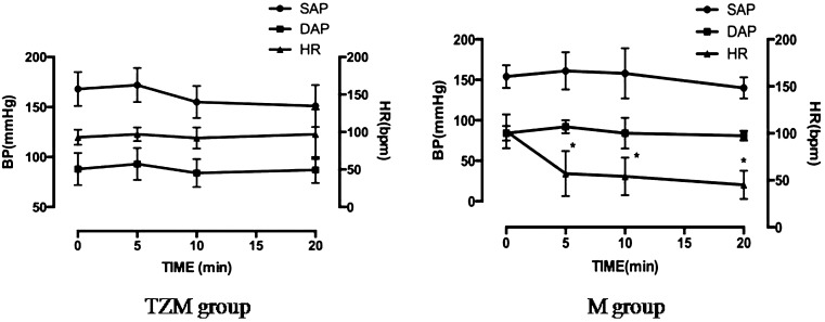
Contrast enhanced ultrasound (CEUS) is useful to evaluate tissue perfusion in the kidney. In veterinary medicine, sedation or anesthesia may be required in uncooperative or panting patients. The aim of this study was to evaluate and compare the normal kidney perfusion patterns in conscious and anesthetized dogs using CEUS. Eight healthy beagles were used in this study. Scanning was performed in conscious dogs using manual restraint (conscious group), or under general anesthesia using tiletamine-zolazepam and medetomidine (TZM group) or medetomidine (M group). The contrast agent (Sonovue(®)) was administered as an IV bolus. The peak intensity (PI), time to peak enhancement from injection (TTP0) and the time to peak enhancement from the initial rise (TTPup), upslope, downslope and area under the curve (AUC) were analyzed. Compared to the cortical values in the conscious group, TTP0 was significantly delayed in the TZM group, and upslope, TTP0 and TTPup were significantly different in the M group. The AUCs in the TZM and M groups were not different from those in the conscious group. The upslope of renal medullary perfusion was significantly decreased in the TZM and M groups. TTP0 and TTPup were also significantly delayed in these groups. The AUC of the medulla was significantly decreased in the M group. Therefore, TZM is useful as an anesthetic protocol when performing CEUS, and the obtained data may serve as reference values in the evaluation of renal perfusion using CEUS in dogs under anesthesia.


Figures

Fig. 1.
Fig. 1.
Scheme of a time-intensity curve with contrast enhanced ultrasonography parameters from a representative dog. The x-axis represents time in seconds and the y-axis represents intensity in decibels. The following functional parameters were derived from the curve: PI (dB) indicates peak intensity; TTP0 (sec) is the time to peak enhancement from the time of contrast agent injection; TTPup (sec) is the time to peak enhancement from the time when the contrast level is initially above baseline; upslope (dB/sec) and downslope (dB/sec) are regression lines of the time-intensity curve between 10% above baseline to 85% of the PI.
Fig. 2.
Fig. 2.
Contrast enhanced images of the left kidney in different stages after a bolus injection of contrast agent. (A) 6 sec after the injection; base line; (B) 9 sec: the contrast agent is diffusely spread in the cortex; (C) 11 sec: the cortical enhancement has reached its peak; the medulla is not enhanced; (D) 15 sec: the contrast agent in the cortex is starting to fade; (E) 22 sec: the medullary perfusion is at its peak; (F) 70 sec: the contrast agent is fully washed out of the cortex and medulla.
Fig. 3.
Fig. 3.
Time-intensity curves obtained from three representative dogs from the A) conscious group, B) TZM group and C) M group.
Fig. 4.
Fig. 4.
Blood pressure and heart rate following administration of anesthetics in the tiletamine-zolazepam and medetomidine (TZM) and metetomidine (M) groups.

References

    1. Bahr A., Wrigley R., Salman M.2000. Quantitative evaluation of IMAGENT® as an abdominal ultrasound contrast medium in dogs. Vet. Radiol. Ultrasound 41: 50–55. doi: 10.1111/j.1740-8261.2000.tb00427.x - DOI - PubMed
    1. Chang J., Kim S., Jung J., Lee H., Choi H., Chang D., Lee Y., Yoon J., Choi M.2011. Assessment of glomerular filtration rate with dynamic computed tomography in normal Beagle dogs. J. Vet. Sci. 12: 393–399. doi: 10.4142/jvs.2011.12.4.393 - DOI - PMC - PubMed
    1. Cullen L. K.1996. Medetomidine sedation in dogs and cats: a review of its pharmacology, antagonism and dose. Br. Vet. J. 152: 519–535. doi: 10.1016/S0007-1935(96)80005-4 - DOI - PubMed
    1. Dong Y., Wang W., Cao J., Fan P., Lin X.2013. Quantitative evaluation of contrast-enhanced ultrasonography in the diagnosis of chronic ischemic renal disease in a dog model. PLoS ONE 8: e70337. doi: 10.1371/journal.pone.0070337 - DOI - PMC - PubMed
    1. Gruenewald S. M., Huster T., Larcos G., Farlow D. C., Choong K. K., Mudaliar Y.1999. Acute renal failure in critically ill patients: evaluation of an ultrasound contrast agent. Australas. Radiol. 43: 180–184. doi: 10.1046/j.1440-1673.1999.00655.x - DOI - PubMed

Publication types

LinkOut - more resources