. 2015 Sep 28:5:14284.
doi: 10.1038/srep14284.

Jagged-2 enhances immunomodulatory activity in adipose derived mesenchymal stem cells

Affiliations

Jagged-2 enhances immunomodulatory activity in adipose derived mesenchymal stem cells

Zhu Xishan et al. Sci Rep. .

Abstract

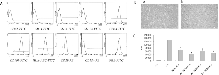
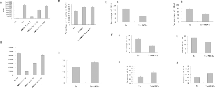
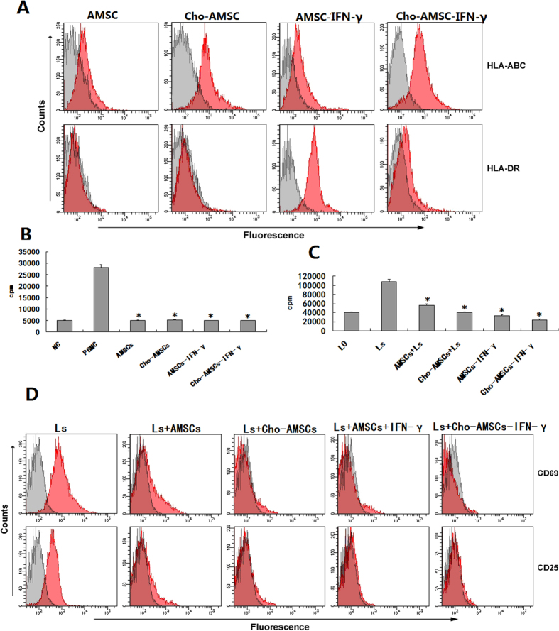
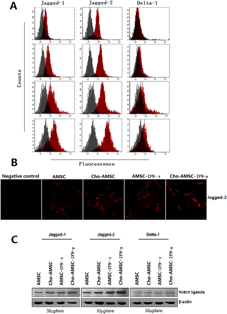
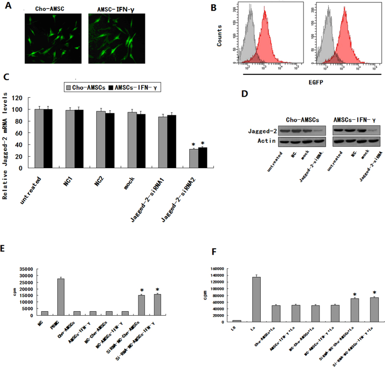
Adipose derived Mesenchymal stem cells (AMSCs) are able to expand in vitro and undergo differentiation into multiple cell lineages, yet have low immunogenicity while exhibiting several immunoregulatory characteristics. We sought to investigate the immunomodulatory mechanisms of AMSCs to better understand their immunogenic properties. Following 10 days of chondrogenic differentiation or 48 hours of IFN-γ pretreatment, AMSCs retained low level immunogenicity but prominent immunoregulatory activity and AMSC immunogenicity was enhanced by chondrogenic differentiation or IFN-γ treatment. We found Jagged-2 expression was significantly elevated following chondrogenic differentiation or IFN-γ pretreatment. Jagged-2-RNA interference experiments suggested that Jagged-2-siRNA2 suppresses Jagged-2 expression during chondrogenic differentiation and in IFN-γ pretreated AMSCs. Besides, Jagged-2 interference attenuated immunosuppressive activity by mixed lymphocyte culture and mitogen stimulation experiments. So, the immunoregulatory activity of AMSCs, to some extent dependent upon Jagged-2, might be stronger after multilineage differentiation or influence from inflammatory factors. This may also be why rejection does not occur after allogeneic AMSCs differentiate into committed cells.


Figures

Figure 1
Figure 1. Biological characteristics of AMSCs.
(A) The morphology of AMSCs (a: before Jagged-2 RNAi; b:after Jagged-2 RNAi) (magnification × 100). (B). Isotype analysis showed cells were all consistently negative for CD34, Sca-1, and CD31 but positive for Flk1, CD29, CD44, and CD105. (C). AMSCs cultured in osteogenic, adipogenic or angiogenic media continuously, sustain their immunoregulatory activities. AMSCs (1 × 104/well) were irradiated by 30 Gy after bone, adipose, and endothelial differentiation and then co-cultured with 50 μg/ml PHA stimulated lymphocytes (1 × 105/well) for 2 days. 0.037 MBq/well 3H-TdR were added and cells incubated for 18 hours. Liquid scintillation was used to determined lymphocyte proliferation.
Figure 2
Figure 2. The effect of AMSCs on T lymphocyte proliferation.
(A) The effects of AMSCs on T lymphocyte proliferation in mitogen proliferation assays. Three groups were assayed: non-stimulated T cells (none), PHA-stimulated T cells (Ts) and PHA-stimulated T cells co-cultured with MSC at different ratios (AMSC to T cell = 1:2, 1:10, 1:100). (B) The effect of AMSCs on T lymphocyte proliferation in MLR. AMSCs at a 1:10 ratio (irradiated MSCs to T cells). (C) Effects of AMSCs on T lymphocyte cell cycle. AMSCs or 3T3 at 1:10 ratios (MSCs to T cells). Cell cycle of PHA-stimulated T cells was analyzed for T cells alone (Ts), and co-cultured with AMSCs (aMSC + Ts). The 3T3 cell line was used as control (3T3 + Ts). Data are shown as mean±SD of five independent experiments. (D) Effect of AMSCs on T lymphocyte activation. AMSCs at 1:10 ratios (MSCs to T cells). T cell activation markers CD25, CD69, and CD44 were examined in T cells alone (Ts) and AMSCs co-cultured with activated T cells (AMSCs + Ts). (E) Effect of AMSCs on T cell apoptosis. AMSCs at a 1:10 ratio (AMSCs to T cells). (F) Effects of AMSCs on T cytokine secretion. The results showed that AMSCs could inhibit IL-2 and IFN-γ but had no significant effect on IL-10 and IL-4. These results indicate that under normal immune circumstances, AMSCs may inhibit Th0 to Th1 differentiation.
Figure 3
Figure 3. Immunological analysis of AMSCs after 7 days of chondrogenic differentiation or 48 hours of IFN-γ pretreatment.
(A) FACS analysis for HLA expression in AMSCs following 7 days of chondrogenic differentiation or 48 hours of IFN-γ pretreatment. (B) AMSCs do not elicit a proliferative response from allogeneic lymphocytes after chondrogenic differentiation and/or IFN-γ pretreatment. (C) Inhibition of mitogen-stimulated lymphocyte proliferation by AMSCs increased after chondrogenic and/or IFN-γ pretreatment. (D) Suppression of mitogen-stimulated lymphocyte activity by AMSCs is retained after osteogenesis and/or IFN-γ pretreatment.
Figure 4
Figure 4. Chondrogenic differentiated AMSCs secrete more IL-10, but less TGF-β after IFN-γ pretreatment.
(A) ELISA was used to detect IL-10 in supernatants from MSCs, Cho-MSCs, MSCsIFN-gamma, and Cho-MSCsIFN-gamma. (B) ELISA was used to detect TGF-β in supernatants from MSCs, Cho-MSCs, MSCsIFN-gamma, and Cho-MSCsIFN-gamma.
Figure 5
Figure 5. Chondrogenic differentiated and/or IFN-γ pretreated MSCs up-regulate Jagged-1.
(A) FACS analysis of Notch ligand expression.(B) Indirect immunofluorescence staining detects Jagged-2. (C) Western blot analysis detects Notch ligand expression.
Figure 6
Figure 6. Immunologic characteristics of chondrogenic differentiated and/or IFN-γ pretreated AMSCs are dependent on up-regulation of Jagged-2.
Jagged-2 mRNAi transfection efficiency of chondrogenic differentiated and/or IFN-γ pretreated AMSCs was measured using several methods (A–D). (A) Fluorescent microscopy. (B) FACS. (C) Real-time PCR. (D) Western blot. (E) The proliferation of allogeneic lymphocytes stimulated in MLC experiments of chondrogenic differentiated and/or IFN-γ pretreated AMSCs was enhanced after Jagged-2 mRNAi transfection (*P < 0.05). (F) Chondrogenic differentiated and/or IFN-γ pretreated AMSC proliferation was inhibited following PHA stimulation of lymphocytes (*P < 0.05).

References

    1. Bouville A. et al. Dose reconstruction for the million worker study: status and guidelines. Health Phys. 108, 206–20 (2015). - PMC - PubMed
    1. Esposito M. & Kang Y. Targeting tumor-stromal interactions in bone metastasis. Pharmacol Ther. 141, 222–33 (2014). - PMC - PubMed
    1. Sun Z. et al. Aberrant CpG islands’ hypermethylation of ABCB1 in mesenchymal stem cells of patients with steroid-associated osteonecrosis. J Rheumatol. 40, 1913–20 (2014). - PubMed
    1. Shin P., Msc J. S. & Mauery D. R. The role of community health centers in providing behavioral health care. J Behav Health Serv Res. 40, 488–96 (2014). - PubMed
    1. Holmes B., Castro N. J., Li J., Keidar M. & Zhang L. G. Enhanced human bone marrow mesenchymal stem cell functions in novel 3D cartilage scaffolds with hydrogen treated multi-walled carbon nanotubes. Nanotechnology. 24, 365–371 (2013). - PubMed

Publication types

MeSH terms

LinkOut - more resources