

Accessory scrotum with perineal lipoma diagnosed prenatally: case report and review of the literature
- PMID: 26412897
- PMCID: PMC4574338
Accessory scrotum with perineal lipoma diagnosed prenatally: case report and review of the literature
Abstract
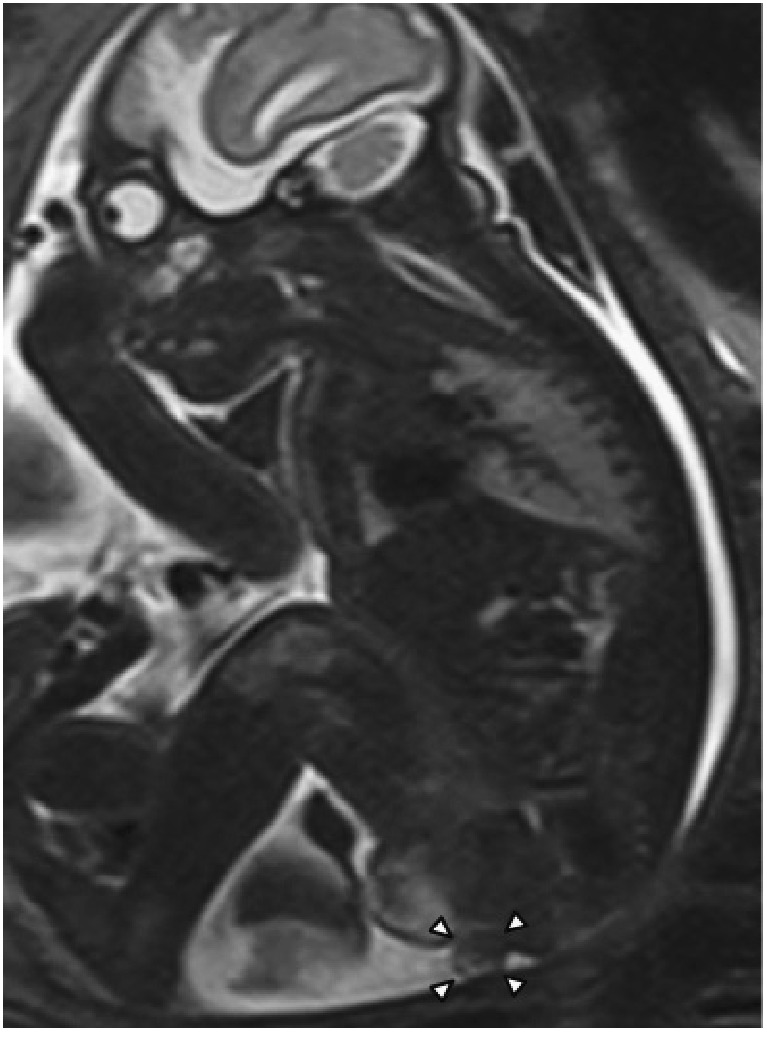
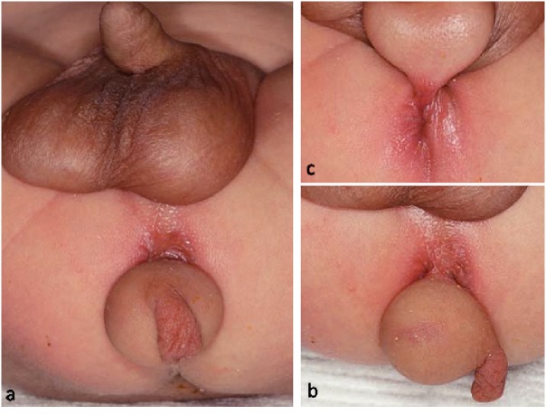
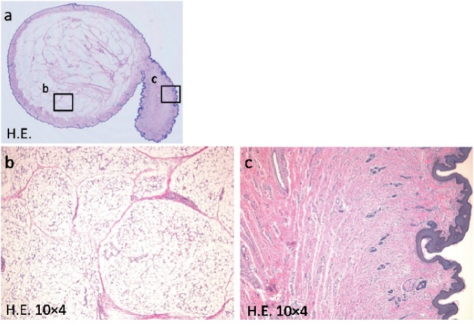
We report a case of accessory scrotum (AS) in the perineal region with peduncular lipoma, diagnosed prenatally. A male fetus of 31 weeks' gestation was referred to our department with a perineal mass. Prenatal ultrasonography and magnetic resonance imaging showed a mass of 1.0 × 1.2 cm located posterior to the scrotum. No other abnormalities were noted during pregnancy. The patient was delivered vaginally at 38 weeks of gestation. On physical examination, a soft peduncular mass with a rugged and pigmented swelling was located between the normally developed scrotum and the anus. There were no specific symptoms or any other associated congenital anomalies. We completely excised the mass at one month of age. A histological examination revealed lipoma, with tissue suggestive of scrotum, so a definite diagnosis of AS was made. AS is a rare congenital anomaly of the scrotum. We review the literature.
Keywords: accessory scrotum; congenital scrotal anomaly; neonate; perineal lipoma; prenatal diagnosis.
Figures
References
-
- Kavecan II, Jovanovic-Privrodski JD, Dobanovacki DS, Obrenovic MR. Accessory scrotum attached to a peduncular perineal lipoma. Pediatr Dermatol, 2012; 29: 522–524. - PubMed
LinkOut - more resources
Full Text Sources
Research Materials