

Abdominal Wall Endometriosis on the Right Port Site After Laparoscopy: Case Report and Literature Review
- PMID: 26412997
- PMCID: PMC4569157
Abdominal Wall Endometriosis on the Right Port Site After Laparoscopy: Case Report and Literature Review
Abstract
Background: Endometriosis can be intrapelvic or, rarely, extrapelvic. Endometriosis involving the rectus abdominis muscle on the trocar port site is a rare event; until now, only 16 cases have been reported in the literature. The majority of cases were associated with previous abdominal surgery such as diagnostic laparoscopy, cyst excision, appendectomy, myomectomy, or cholecystectomy. We review all the reported cases of this unusual form of extrapelvic endometriosis.
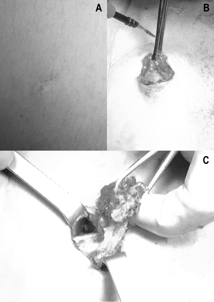
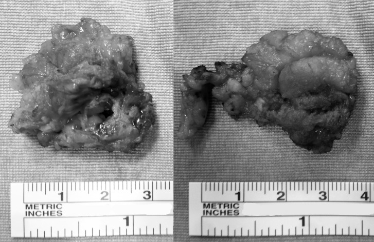
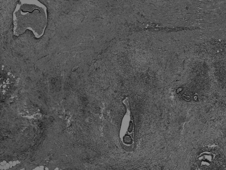
Case report: We report a new case of abdominal wall endometriosis at the trocar port site in the rectus abdominis muscle in a woman who had undergone 2 laparoscopies for endometriosis in the 3 years before coming to our attention. The diagnosis was made by sonography. We performed a surgical resection of the lesion with a free macroscopic margin of 5-10 mm.
Conclusion: Endometriosis should be considered in the differential diagnosis of any abdominal swelling. In our experience, surgery is the treatment of choice.
Keywords: Abdominal wall; endometriosis; laparoscopy.
Figures
References
-
- Burney RO, Giudice LC. Pathogenesis and pathophysiology of endometriosis. Fertil Steril. 2012 Sep;98(3):511–519. doi: 10.1016/j.fertnstert.2012.06.029. - DOI - PMC - PubMed
-
- Eskenazi B, Warner ML. Epidemiology of endometriosis. Obstet Gynecol Clin North Am. 1997 Jun;24(2):235–258. - PubMed
-
- Calò PG, Ambu R, Medas F, Longheu A, Pisano G, Nicolosi A. Rectus abdominis muscle endometriosis Report of two cases and review of the literature. Ann Ital Chir. 2012 Jun 20;pii: [Epub ahead of print] - PubMed
-
- Dordevic M, Jovanovic B, Mitrovic S, Dordevic G, Radovanovic D, Sazdanovic P. Abdominal rectus muscle endometriosis after Cesarean section. Extrapelvic localization of endometriosis. Bratisl Lek Listy. 2010;111(6):345–348. - PubMed
-
- Savelli L, Manuzzi L, Di Donato N, et al. Endometriosis of the abdominal wall: ultrasonographic and Doppler characteristics. Ultrasound Obstet Gynecol. 2012 Mar;39(3):336–340. doi: 10.1002/uog.10052. - DOI - PubMed
LinkOut - more resources
Full Text Sources