Noninfiltrating Adenocarcinoma of the Lung Causing ST-Segment Elevation
- PMID: 26413024
- PMCID: PMC4567106
- DOI: 10.14503/THIJ-14-4268
Noninfiltrating Adenocarcinoma of the Lung Causing ST-Segment Elevation
Abstract
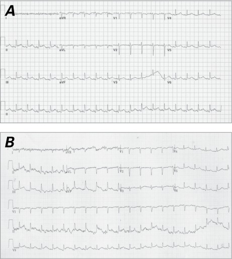
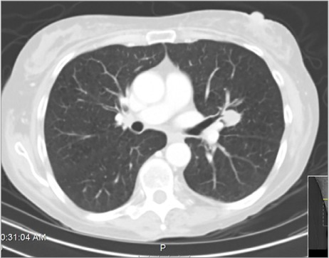
ST-segment-elevation myocardial infarction is a leading cause of cardiovascular morbidity and death. We describe the case of a 51-year-old woman with advanced adenocarcinoma of the lung who presented with ST-segment elevation in the presence of an extracardiac lung mass but no objective evidence of myocardial ischemia or pericardial involvement. After the patient died of hypoxic respiratory failure, autopsy findings confirmed normal-appearing pericardium and myocardium, and mild-to-moderate atherosclerosis in the coronary arteries. A 4.5 × 4-cm extracardiac left hilar lung mass was confirmed to be poorly differentiated adenocarcinoma of the lung adjacent to the myocardium. The persistent current of injury that had been detected electrocardiographically was thought to occur from direct myocardial compression. ST-segment elevations secondary to direct mass contact on the myocardium should be considered in patients who have a malignancy and ST-segment elevation.
Keywords: Arrhythmias, cardiac/diagnosis; diagnosis, differential; electrocardiography; lung neoplasms/complications/pathology; myocardial infarction/classification.
Figures


References
-
- Cooper R, Cutler J, Desvigne-Nickens P, Fortmann SP, Friedman L, Havlik R et al. Trends and disparities in coronary heart disease, stroke, and other cardiovascular diseases in the United States: findings of the national conference on cardiovascular disease prevention. Circulation. 2000;102(25):3137–47. - PubMed
-
- Thygesen K, Alpert JS, Jaffe AS, Simoons ML, Chaitman BR, White HD et al. Third universal definition of myocardial infarction. J Am Coll Cardiol. 2012;60(16):1581–98. - PubMed
-
- O'Gara PT, Kushner FG, Ascheim DD, Casey DE, Jr, Chung MK, de Lemos JA et al. 2013 ACCF/AHA guideline for the management of ST-elevation myocardial infarction: a report of the American College of Cardiology Foundation/American Heart Association Task Force on Practice Guidelines [published erratum appears in Circulation 2013;128(25):e481] Circulation. 2013;127(4):e362–425. - PubMed
-
- Wang K, Asinger RW, Marriott HJ. ST-segment elevation in conditions other than acute myocardial infarction. N Engl J Med. 2003;349(22):2128–35. - PubMed
-
- Astorri E, Fiorina P, Pattoneri P, Paganelli C. Persistent ST-segment elevation in a patient with metastatic involvement of the heart. Minerva Cardioangiol. 2001;49(1):81–5. - PubMed
Publication types
MeSH terms
LinkOut - more resources
Full Text Sources
Medical