Percutaneous Management of a Coronary Bifurcation Aneurysm with Mesh-Covered Stents and the Simultaneous Kissing Stent Technique
- PMID: 26413028
- PMCID: PMC4567121
- DOI: 10.14503/THIJ-13-4020
Percutaneous Management of a Coronary Bifurcation Aneurysm with Mesh-Covered Stents and the Simultaneous Kissing Stent Technique
Abstract
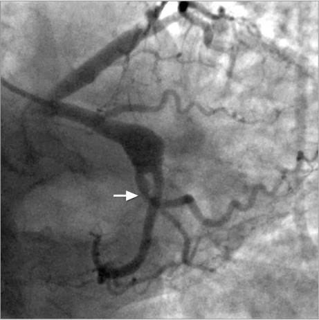
A 63-year-old man was admitted with a clinical diagnosis of acute coronary syndrome (non-ST-segment elevation), characterized by regional hypokinesia of the left ventricular posterior and lateral walls and by positive cardiac biomarkers. The coronary angiogram showed a 12.5-mm-diameter aneurysm with a mural thrombus and possible distal embolism to the bifurcation of the left circumflex coronary artery and the 2nd marginal branch. The aneurysm was managed percutaneously by implanting 2 mesh-covered stents in accordance with the "simultaneous kissing stent" technique. Follow-up angiography and optical coherence tomography at 5 postprocedural months documented complete sealing of the aneurysm and diffuse in-stent restenosis. No sign of ischemia occurred during the subsequent follow-up.
Keywords: Acute coronary syndrome; bifurcation aneurysm; blood vessel prosthesis implantation; coronary aneurysm/diagnosis/therapy; disease progression; percutaneous coronary intervention/methods; stents.
Figures


References
-
- Crimi G, Bellotti S, Iannone A, Rubartelli P. Sequential coronary angiograms unveil the progression of an acquired coronary aneurysm. Eur Heart J. 2013;34(37):2924. - PubMed
-
- Syed M, Leach M. Coronary artery aneurysm: a review. Prog Cardiovasc Dis. 1997;40(1):77–84. - PubMed
-
- Gunduz H, Akdemir R, Binak E, Tamer A, Uyan C. Spontaneous rupture of a coronary artery aneurysm: a case report and review of the literature. Jpn Heart J. 2004;45(2):331–6. - PubMed
-
- Iakovou I, Colombo A. Treatment of a coronary aneurysm involving bifurcation with the use of a custom-made polytetrafluoroethylene-covered bifurcation stent system. Catheter Cardiovasc Interv. 2005;64(2):169–72. - PubMed
Publication types
MeSH terms
LinkOut - more resources
Full Text Sources