. 2015:2015:810796.
doi: 10.1155/2015/810796. Epub 2015 Aug 27.

Fast and Accurate Semiautomatic Segmentation of Individual Teeth from Dental CT Images

Affiliations

Fast and Accurate Semiautomatic Segmentation of Individual Teeth from Dental CT Images

Ho Chul Kang et al. Comput Math Methods Med. 2015.

Abstract

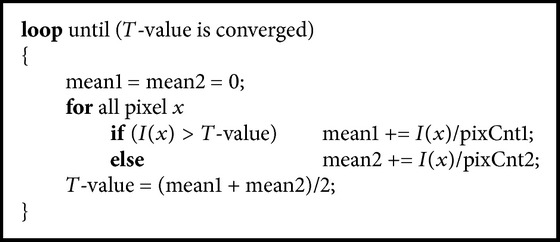
In this paper, we propose a fast and accurate semiautomatic method to effectively distinguish individual teeth from the sockets of teeth in dental CT images. Parameter values of thresholding and shapes of the teeth are propagated to the neighboring slice, based on the separated teeth from reference images. After the propagation of threshold values and shapes of the teeth, the histogram of the current slice was analyzed. The individual teeth are automatically separated and segmented by using seeded region growing. Then, the newly generated separation information is iteratively propagated to the neighboring slice. Our method was validated by ten sets of dental CT scans, and the results were compared with the manually segmented result and conventional methods. The average error of absolute value of volume measurement was 2.29 ± 0.56%, which was more accurate than conventional methods. Boosting up the speed with the multicore processors was shown to be 2.4 times faster than a single core processor. The proposed method identified the individual teeth accurately, demonstrating that it can give dentists substantial assistance during dental surgery.


Figures

Figure 1
Figure 1
The axial view of the human mandible which is extracted with a threshold value: (a) is the original image of CT and (b) is the magnified view of the original image.
Figure 2
Figure 2
The histogram of CT scans of the mandible. It shows the dark background, tissues, and bones in the order from the darkest to the lightest.
Figure 3
Figure 3
The examples of teeth images. The difference in the brightness between teeth and sockets of teeth in dental CT images is relatively low.
Figure 4
Figure 4
An overview of the proposed segmentation of individual teeth. First, we reduce noise and set T-value. Second, we fill holes of teeth and remove the branches. Finally, we check that the result of segmentation is oversegmented or undersegmented. If the result is over or under, go back to T-value step.
Figure 5
Figure 5
Initial T-value. This should be easily classified as the low density of tissues and the high density of bones.
Figure 6
Figure 6
The bisection search: optimal T-value preventing the oversegmentation while keeping the shape and size of segmented teeth from the previous slice.
Figure 7
Figure 7
A memory access order for improving the cache efficiency. If we explored the right side, we can improve the efficiency by 5 to 10 times.
Figure 8
Figure 8
Unexpected holes shown by dentin and pulp while using simple SRG.
Figure 9
Figure 9
Filling the holes by SRG. We set the bounding box and fill up holes using inverse-SRG and complement operator.
Figure 10
Figure 10
The result of teeth segmentation after filling up empty spaces.
Figure 11
Figure 11
Resetting the oversegmented regions compared to the segmented region of the previous slice.
Figure 12
Figure 12
To minimize memory access conflicts among threads in the region growing algorithm, the initial seed points are spread in a 3D way.
Figure 13
Figure 13
Results of segmented individual teeth by proposed method: isolated individual teeth are marked in red.
Figure 14
Figure 14
Result of threshold techniques after altering threshold values: (a) is the axial view of the original; (b) and (c) are the results of threshold techniques altering threshold values, which cause under- or oversegmentation.
Pseudocode 1
Pseudocode 1
Pseudocode 2
Pseudocode 2

References

    1. Mathew C. A., Maller S., Maheshwaran Interactions between magnetic resonance imaging and dental material. Journal of Pharmacy & Bioallied Sciences. 2013;5(1):S113–S116. doi: 10.4103/0975-7406.113309. - DOI - PMC - PubMed
    1. Verstreken K., van Cleynenbreugel J., Martens K., Marchai G., van Steenberghe D., Suetens P. An image-guided planning system for endosseous oral implants. IEEE Transactions on Medical Imaging. 1998;17(5):842–852. doi: 10.1109/42.736056. - DOI - PubMed
    1. Misch C. E. Contemporary implant dentistry. Implant Dentistry. 1999;8(1):p. 90.
    1. Futterling S., Klein R., Straber W., Weber H. Automated finite element modeling of a human mandible with dental implants. Proceedings of the 6th International Conference in Central Europe on Computer Graphics and Visualization; 1998; pp. 103–110.
    1. Rueda S., Gil J. A., Pichery R., Alcañiz M. Automatic segmentation of jaw tissues in CT using active appearance models and semi-automatic landmarking. Medical Image Computing and Computer-Assisted Intervention. 2006;9, part 1:167–174. - PubMed

Publication types

MeSH terms