. 2014 Jul 1:7:100.
doi: 10.1186/1754-6834-7-100. eCollection 2014.

Cellulosomics of the cellulolytic thermophile Clostridium clariflavum

Affiliations

Cellulosomics of the cellulolytic thermophile Clostridium clariflavum

Lior Artzi et al. Biotechnol Biofuels. .

Abstract

Background: Clostridium clariflavum is an anaerobic, thermophilic, Gram-positive bacterium, capable of growth on crystalline cellulose as a single carbon source. The genome of C. clariflavum has been sequenced to completion, and numerous cellulosomal genes were identified, including putative scaffoldin and enzyme subunits.

Results: Bioinformatic analysis of the C. clariflavum genome revealed 49 cohesin modules distributed on 13 different scaffoldins and 79 dockerin-containing proteins, suggesting an abundance of putative cellulosome assemblies. The 13-scaffoldin system of C. clariflavum is highly reminiscent of the proposed cellulosome system of Acetivibrio cellulolyticus. Analysis of the C. clariflavum type I dockerin sequences indicated a very high level of conservation, wherein the putative recognition residues are remarkably similar to those of A. cellulolyticus. The numerous interactions among the cellulosomal components were elucidated using a standardized affinity ELISA-based fusion-protein system. The results revealed a rather simplistic recognition pattern of cohesin-dockerin interaction, whereby the type I and type II cohesins generally recognized the dockerins of the same type. The anticipated exception to this rule was the type I dockerin of the ScaB adaptor scaffoldin which bound selectively to the type I cohesins of ScaC and ScaJ.

Conclusions: The findings reveal an intricate picture of predicted cellulosome assemblies in C. clariflavum. The network of cohesin-dockerin pairs provides a thermophilic alternative to those of C. thermocellum and a basis for subsequent utilization of the C. clariflavum cellulosomal system for biotechnological application.

Keywords: Biofuels; Biomass degradation; CBM; Cellulases; Cellulosomes; Cohesin; Dockerin; Glycoside hydrolases; Scaffoldin.


Figures

Figure 1
Figure 1
Pictograms showing modular arrangement of putative scaffoldins of the C. clariflavum DSM 19732 genome. Thirteen putative scaffoldins were identified bioinformatically. Black dots indicate cohesin and dockerin modules of the designated scaffoldins that were expressed and examined for specific interactions in the current study. All sequences contain an N-terminal signal peptide except ScaO and ScaM(a). CBM, carbohydrate-binding module; CSBM, cell surface-binding module; FN3, fibronectin type III domain; CARDB, cell adhesion-related domain found in bacteria; DUF11, domain of unknown function (Pfam PF01345); BIL, bacterial intein-like domain; SLH, S-layer homology. Accession numbers of C. clariflavum scaffoldins: [YP_005047733 (ScaA), YP_005047732 (ScaB), YP_005047731.1 (ScaC), YP_005047730 (ScaD), YP_005046332 (ScaE), YP_005047223 (ScaF), YP_005046504 (ScaG), YP_005047817 (ScaH/L), YP_005047757 (ScaJ), YP_005048513 (ScaM), YP_005048561 (ScaM(a)), YP_005048562 (ScaM(b)), YP_005046147 (ScaO): GeneBank].
Figure 2
Figure 2
Phylogeny of C. clariflavum cohesins. A set of 47 C. clariflavum (Cc), 41 A. cellulolyticus (Ac), and 18 C. thermocellum (Ct) cohesin-like modules, derived from deduced amino-acid sequences (supporting Additional file 1: Figure S1), was aligned using the CLUSTALW2 program at the EBI website [35], which then served to reconstruct an unrooted phylogenetic tree by the MEGA5.10 software [36], using the neighbor-joining method with 500 bootstrap replicates. Numerical values above the nodes indicate bootstrap percentiles. The cohesin-like modules distribute into two major classes: type I (yellow) and type II (green). Among the type I cohesin-like modules one subgroup is separated from the majority of the modules (pink).
Figure 3
Figure 3
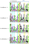
Comparative sequence logos of the C. clariflavum and A. cellulolyticus dockerin modules. Amino acid conservation of the type I dockerin repeat sequences was performed by a logo, created using WebLogo (see Methods) based on 74 type I dockerin sequences of C. clariflavum and 138 of A. cellulolyticus. The top logo of each represents the first dockerin sequence repeat and the bottom logo represents the second dockerin repeat. Calcium-binding residues are highlighted in light blue, and the presumed recognition residues responsible for cohesin-dockerin interactions are highlighted in yellow.
Figure 4
Figure 4
Sequences of C. clariflavum and A. cellulolyticus ScaB dockerins. Sequence alignment of the two dockerin modules was performed using the CLUSTALW2 program at the EBI website. Consensus residues are as defined accordingly; *indicates a position which has an identical residue, and colon (:) and period (.) indicate conservation between groups of strongly and weakly similar properties, respectively; blue indicates conservation between species and green indicates conservation between the two repeated segments. Ca+2-binding residues are highlighted in cyan, and putative recognition residues are highlighted in yellow. Residues are numbered relative to the highly conserved glycine (designated 0), which is positioned adjacent to the initial calcium-binding aspartate (residue 1).
Figure 5
Figure 5
Determination of cohesin-dockerin specificity by affinity-based ELISA. In order to identify specific interactions between the nineteen CBM-Cohs and the four XynDocs, microtiter plates were coated with the respective CBM-Coh fusion protein, and increasing concentrations of the various XynDocs were then applied to the plates. The EC50 was calculated for the resultant interactions, and values of the pEC50 are presented on the y-axis in the bar graph. Coh, cohesin; Doc, dockerin; XDoc, X-dockerin modular dyad; CBM, carbohydrate-binding module. The cohesin names and numbers are shown on the horizontal axis (for example, A1 indicates the first cohesin of ScaA). Xyn-XDocA, green; Xyn-XDocH/L, dark green; XynDocB, red; XynDocGH48, yellow.
Figure 6
Figure 6
Proposed architectures for cell-bound and cell-free cellulosome assembly in C. clariflavum. The scheme shows the possible interactions among scaffoldin and enzymatic modules, as derived from examination of interactions by affinity ELISA. Specification of scaffoldins is detailed in Figure 1. Four potential cell-anchored cellulosomal complexes are represented. Two of the complexes employ the adaptor protein ScaB to join the cell-anchored scaffoldins (ScaC and ScaJ, containing an SLH domain, and four and one type I cohesins, respectively) to the primary enzyme-integrating scaffoldins (ScaA and ScaH/L) via the type II cohesins of ScaB and XDocs of the former. The type II cohesins of ScaD (cohesins 1 and 2) and ScaF are also cell-anchored scaffoldins that bind directly ScaA or ScaH/L. The type I cohesins of ScaG and ScaD (cohesin 3) interact with type I dockerins of dockerin-bearing enzymes. ScaG is suspected to be a cell-anchored scaffoldin, based on previous studies of the copper amine-oxidase domain in the OlpC protein from C. thermocellum. ScaE has seven type II cohesins which are able to bind seven XDoc modules, thereby creating a large, cell-free cellulosomal complex. CBSM, cell surface-binding module.

References

    1. Bayer EA, Lamed R. The cellulose paradox: pollutant par excellence and/or a reclaimable natural resource? Biodegradation. 1992;3:171–188. - PubMed
    1. Bayer EA, Lamed R, Himmel ME. The potential of cellulases and cellulosomes for cellulosic waste management. Curr Opinion Biotechnol. 2007;18:237–245. - PubMed
    1. Gilbert HJ. The biochemistry and structural biology of plant cell wall deconstruction. Plant Physiol. 2010;153:444–455. - PMC - PubMed
    1. Atalla RH. In: Comprehensive natural products chemistry. Pinto BM, editor. Volume 3. Cambridge: Elsevier; 1999. Celluloses; pp. 529–598.
    1. O'Sullivan AC. Cellulose: the structure slowly unravels. Cellulose. 1997;4:173–207.

LinkOut - more resources