Review
. 2014 Dec 31;4(2):e979619.
doi: 10.4161/21565562.2014.979619. eCollection 2014 May-Aug.

The blood-epididymis barrier and inflammation

Affiliations
Review

The blood-epididymis barrier and inflammation

Mary Gregory et al. Spermatogenesis. .

Abstract

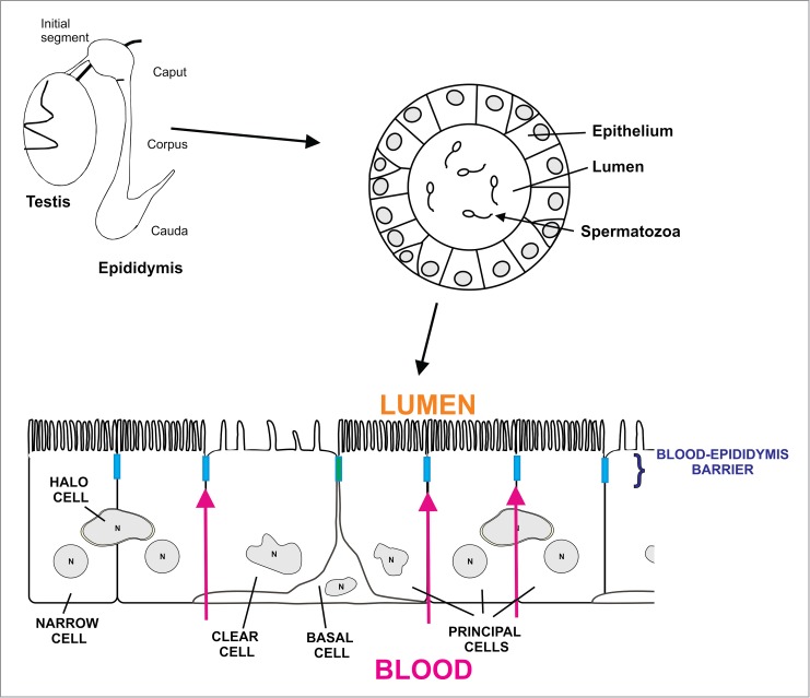
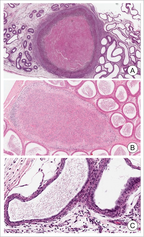
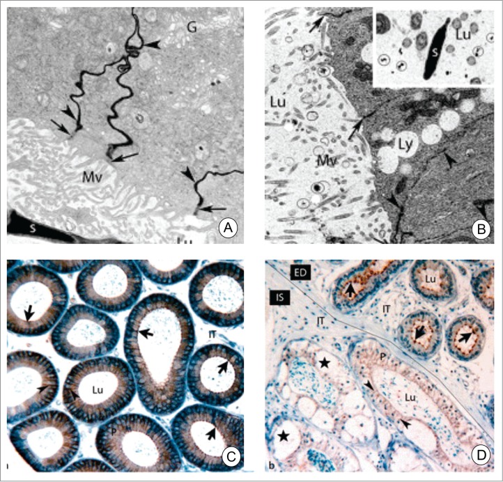
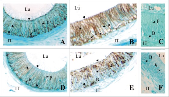
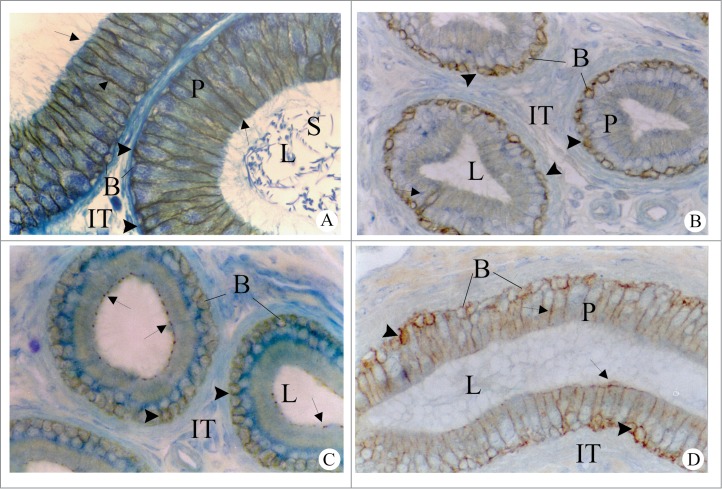
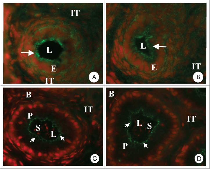
The blood-epididymis barrier (BEB) is a critical structure for male fertility. It enables the development of a specific luminal environment that allows spermatozoa to acquire both the ability to swim and fertilize an ovum. The presence of tight junctions and specific cellular transporters can regulate the composition of the epididymal lumen to favor proper sperm maturation. The BEB is also at the interface between the immune system and sperm. Not only does the BEB protect maturing spermatozoa from the immune system, it is also influenced by cytokines released during inflammation, which can result in the loss of barrier function. Such a loss is associated with an immune response, decreased sperm functions, and appears to be a contributing factor to post-testicular male infertility. Alterations in the BEB may be responsible for the formation of inflammatory conditions such as sperm granulomas. The present review summarizes current knowledge on the morphological, physiological and pathological components associated with the BEB, the role of immune function on the regulation of the BEB, and how disturbance of these factors can result in inflammatory lesions of the epididymis.

Keywords: ABC transporters; claudins; defensin; granuloma; sperm maturation.


Figures

Figure 1.
Figure 1.
Schematic diagram showing the testis and epididymis including the 4 regions of the epididymis (initial segment, caput, corpus and cauda epididymidis). The epididymis is a single convoluted tubule in which the epithelium regulates the composition of the lumen necessary for sperm to acquire the ability to swim and fertilize. The epithelium of the epididymis contains various cells types, including principal cells, clear cells, basal cells and halo cells. Apical and narrow cells are present exclusively in the initial segment. Apical tight junctions between the cells that line the lumen of the epididymis form the blood-epididymis barrier. The barrier regulates the content of the epididymal lumen by limiting paracellular movement of ions between the blood and the lumen. It also protects maturing spermatozoa from the immune system.
Figure 2.
Figure 2.
Epididymal granuloma and inflammation. Examples of epididymal granuloma (panels A and B). Note the alterations and degradation of the epithelium around the granuloma containing accumulated sperm. Mixed immune cells can be observed either surrounding (A) or in the periphery of the granuloma (B). Panel B shows an inflammatory response with increased immune cells in the interstitium. Photomicrographs were generously provided by Dr. D. Creasy (Huntingdon Life Sciences).
Figure 3.
Figure 3.
Electron microscope images of adult wild-type (A and B) and deficient in protective protein cathepsin A mice perfused with lanthanum nitrate. In A, lanthanum nitrate percolates through the intercellular spaces (arrowheads) between adjacent principal cells up to the apical tight junctions. In B, lanthanum nitrate permeates the entire intercellular space (arrowheads) including the apical junctional complexes (arrows) indicating a loss of tight junctions. Lanthanum nitrate is also evident in the epididymal lumen (circles, inset of B). In C, paraffin sections of wild-type mice were immunostained for Cldn3. Cldn3 is located in the apical region of the epithelium (arrow). In cathepsin A-knockout mice, the localization of Cldn3 is cytoplasmic. Furthermore, the interstitial area contains numerous macrophages and large vacuoles are present in the epithelium. Reproduced from Figures 1 and 3 of Hermo et al.
Figure 4.
Figure 4.
Immunolocalization of CLDN1 (A, B) and CLDN3 (C, D) in the caput epididymides of fertile men (A) and infertile men (B). Negative controls with no primary antibodies are shown (C). While CLDN1 and CLDN3 are localized (arrowheads) to the area of tight junctions between adjacent principal cells in the caput epididymidis of fertile patients, in In infertile patients there is a noticeable increase in cytosolic localization. These results show that the localization of these CLDNS can be negatively regulated in infertile men. Magnification × 640. P, Principal cells; B, basal cells; IT, intertubular space; Lu, lumen. Reproduced from Dubé et al..
Figure 5.
Figure 5.
Regulation of Cldn1 in the initial segment (AC) of the adult rat epididymis. In intact adult rats (A), Cldn1 is immunolocalized to the lateral plasma membranes between neighboring principal cells (arrows) and between principal and basal cells (arrowheads). In adult rats, 14 days after orchidectomy (B), immunoreactive Cldn1 is associated at the interface between basal and principal cells (arrowheads) and not between the lateral plasma membranes of adjacent principal cells. In orchidectomized rats given testosterone replacement (C), immunoreactive Cldn1 is observed between the lateral plasma membranes at apical sites in areas of tight junctions (arrows), but not at more distal sites of these membranes; reaction, however, is maintained between basal and principal cells (arrowheads). P, Principal cells; B, basal cells; IT, intertubular space; L, lumen. Magnification, ×640. Reproduced from Gregory et al.,.
Figure 6.
Figure 6.
Immunocytochemical localization of Cldn-3 (A, C) and Cldn-4 (B, D) in the epididymis of 14 (A, B)- and 42 (C, D) day old rats. At 14 days there is an important cytoplasmic reaction (arrows) suggesting that Cldns have not yet completely localized to the area of the tight junction. In 42 day old rats the reaction in all regions of the epididymis is present exclusively in the area of tight junction and there is no cytoplasmic staining. The photomicrographs are from the initial segment region. Magnification 640×. E = epithelial cells, P = principal cells, IT = intertubular space, L = lumen. Reproduced from Gregory et al.,.

References

    1. Hinton BT, Howards SS. Permeability characteristics of the epithelium in the rat caput epididymidis. J Reprod Fertil. 1981; 63:95-9; PMID:7277337; http://dx.doi.org/10.1530/jrf.0.0630095 - DOI - PubMed
    1. Friend DS, Gilula NB. Variations in tight and gap junctions in mammalian tissues. J Cell Biol. 1972; 53:758-76; PMID:4337577; http://dx.doi.org/10.1083/jcb.53.3.758 - DOI - PMC - PubMed
    1. Suzuki F, Nagano T. Regional differentiation of cell junctions in the excurrent duct epithelium of the rat testis as revealed by freeze-fracture. Anat Rec 1978; 191:503-19; PMID:697060; http://dx.doi.org/10.1002/ar.1091910409 - DOI - PubMed
    1. Cyr DG, Robaire B, Hermo L. Structure and turnover of junctional complexes between principal cells of the rat epididymis. Microsc Res Tech 1995; 30:54-66; PMID:7711320; http://dx.doi.org/10.1002/jemt.1070300105 - DOI - PubMed
    1. Agarwal A, Hoffer AP. Ultrastructural studies on the development of the blood-epididymis barrier in immature rats. J Androl 1989; 10:425-31; PMID:2621151 - PubMed