. 2015 Feb 23;5(1):e1006089.
doi: 10.1080/21565562.2015.1006089. eCollection 2015 Jan-Apr.

Drosophila spermatid individualization is sensitive to temperature and fatty acid metabolism

Affiliations

Drosophila spermatid individualization is sensitive to temperature and fatty acid metabolism

Geulah Ben-David et al. Spermatogenesis. .

Abstract

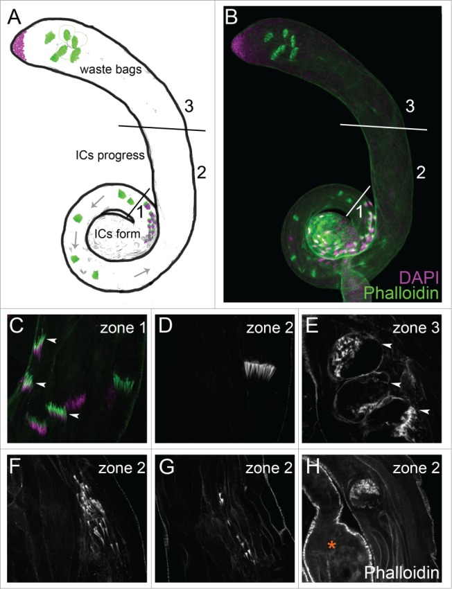
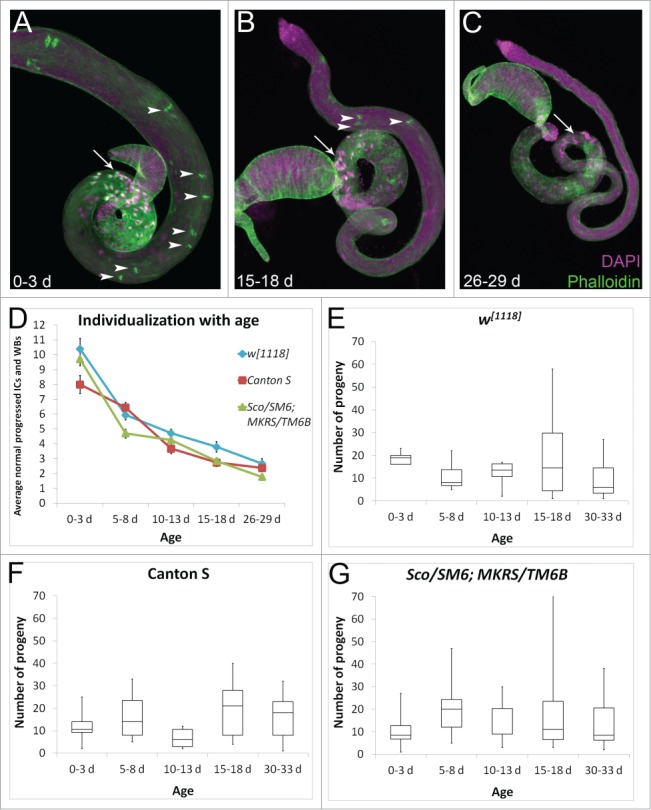
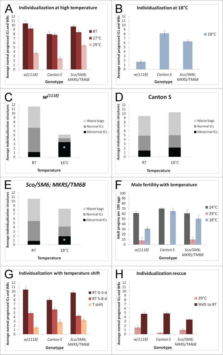
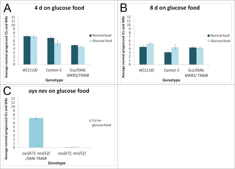
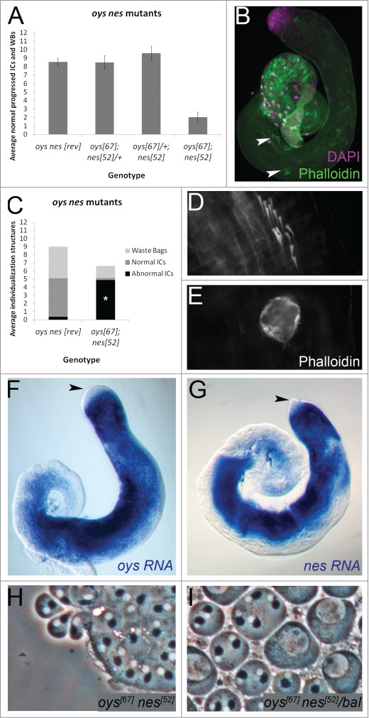
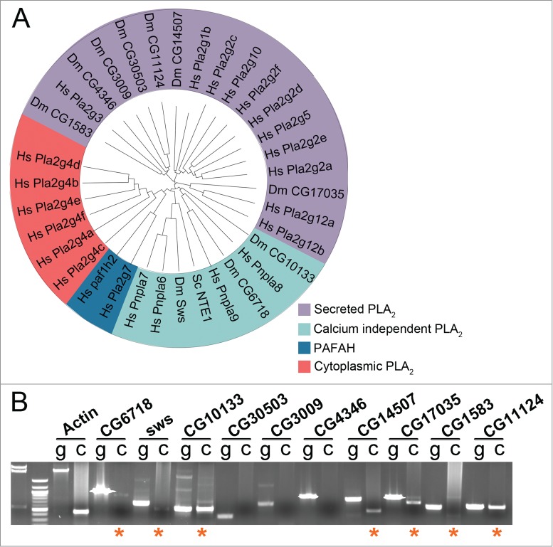
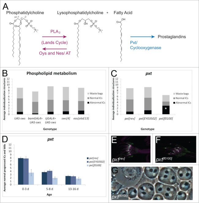
Fatty acids are precursors of potent lipid signaling molecules. They are stored in membrane phospholipids and released by phospholipase A2 (PLA2). Lysophospholipid acyltransferases (ATs) oppose PLA2 by re-esterifying fatty acids into phospholipids, in a biochemical pathway known as the Lands Cycle. Drosophila Lands Cycle ATs oys and nes, as well as 7 predicted PLA2 genes, are expressed in the male reproductive tract. Oys and Nes are required for spermatid individualization. Individualization, which occurs after terminal differentiation, invests each spermatid in its own plasma membrane and removes the bulk of the cytoplasmic contents. We developed a quantitative assay to measure individualization defects. We demonstrate that individualization is sensitive to temperature and age but not to diet. Mutation of the cyclooxygenase Pxt, which metabolizes fatty acids to prostaglandins, also leads to individualization defects. In contrast, modulating phospholipid levels by mutation of the phosphatidylcholine lipase Swiss cheese (Sws) or the ethanolamine kinase Easily shocked (Eas) does not perturb individualization, nor does Sws overexpression. Our results suggest that fatty acid derived signals such as prostaglandins, whose abundance is regulated by the Lands Cycle, are important regulators of spermatogenesis.

Keywords: actin; drosophila; fatty acids; individualization; lipid metabolism; prostaglandins; spermiogenesis.


Figures

Figure 1.
Figure 1.
(See previous page). Characterization of normal and abnormal individualization structures. (A, B) Testes can be divided schematically into 3 domains. In zone 1, the ICs (phalloidin, green) form in apposition to the nuclei (DAPI, purple). The ICs migrate away from the nuclei along the spermatid axonemes toward the apical testis (zone 2). In zone 3, the ICs are degraded within the waste bags. (CE) Testes from Sco/SM6;MKRS/TM6B. (C) Newly formed actin cones in zone 1 are thin and needle-shaped (arrowheads). (D) As the IC progresses, actin cones in zone 2 become conical and migrate synchronously. (E) Waste bags in zone 3 are bounded by cortical actin and contain the degenerating actin cones (arrowheads). (F-H) Testes from Canton S males reared at 29oC. Abnormal ICs contain scattered actin cones (F), unaffiliated actin cones (G), or degenerate before reaching zone 3, as indicated in the photograph by the proximity to the seminal vesicle (asterisk, H).
Figure 2.
Figure 2.
(See previous page). The effects of aging on spermatogenesis. (A) Young testes are large and have many late stage spermatogenic cysts with clustered nuclei in the basal testis (arrow, DAPI, purple) and many progressing ICs (arrowheads, phalloidin, green). (B, C) Older testes are thin and have fewer cysts with basal nuclear clusters (arrows) and fewer ICs (arrowheads). Testes are from w1118 males. (D) Young (0–3 d old) males at room temperature from 3 control genotypes have 8–11 normal progressed ICs and waste bags (WBs) per testis on average. Older males show a decline in the average number of individualization structures per testis, with 5–8 d old males having 4–7 normal individualization structures, 10–13 d old males having 3–5 normal individualization structures, 15–18 d old males having 2–4 normal individualization structures, and 26–29 d old males having fewer than 3 normal individualization structures per testis on average. N ≥ 18 testes each data point. In this an all subsequent figures, error bars represent standard error mean. (EG) Fertility in 0–3 d old, 5–8 d old, 10–13 d old, 15–18 d old, or 30–33 d old males assessed in single matings (one male and one female each) for each control genotype (w1118, Canton S, or the balancer stock Sco/SM6;MKRS/TM6B). F1 progeny from 10 individual crosses were counted. Box plots show the distribution of progeny numbers for each age group, including maximum, minimum, interquartile range, and median numbers of progeny. ANOVA analysis comparing the mean number of progeny in each age group within each genotype reveals p > 0.1 for all 3 genotypes.
Figure 3.
Figure 3.
(See previous page). Individualization is sensitive to temperature. (A) Young (0–3 d old) males raised at 27oC have a slight decrease in the average number of normal individualization structures per testis (red bars, n ≥ 18 each genotype) compared to room temperature (RT) males of the same genotypes (maroon bars, n ≥ 18 each genotype). At 29oC, testes average fewer than 6 normal individualization structures each (pink bars, n > 10 each genotype). (B) At 18oC, young w1118 males (0–6 d old) have fewer than 2 normal individualization structures per testis on average (n = 57). In contrast, Canton S males at 18oC have more than 8 normal individualization structures per testis (n = 37), and balancer males at 18oC have an average of more than 6 normal individualization structures per testis (n = 34). (C) Young w1118 males raised at room temperature have an average of 1.15 abnormal ICs per testis, compared to young w1118 males raised at 18oC, which have an average of 3.42 abnormal ICs per testis. p < 0.001. (D) Young Canton S males raised at room temperature have an average of 1.44 abnormal ICs per testis, compared to young Canton S males raised at 18oC, which have an average of 2.14 abnormal ICs per testis. (E) Young Sco/SM6;MKRS/TM6B males raised at room temperature have an average of 0.875 abnormal ICs per testis, compared to young Sco/SM6;MKRS/TM6B males raised at 18oC, which have 1.94 abnormal ICs per testis. p < 0.001. (F) Young males raised and mated at 29oC or 18oC have reduced fertility compared to young males raised at 24oC. Number of adult progeny resulting from 100 eggs collected from each cross was counted and averaged between 3 trials. Error bars represent SEM. (G) Allowing males to develop at room temperature and shifting them to 29oC within 0–3 d of eclosion significantly reduces the average number of normal individualization structures per testis (peach bars, T shift, n ≥ 15 each genotype). Temperature shifted males were kept at 29oC for 5 d. Control males were 0–3 d old room temperature males (maroon bars, n ≥ 18 each genotype) or 5–8 d old room temperature males (light red bars, n ≥ 18 each genotype). (H) Rearing males at 29oC and shifting them to room temperature within 0–3 d of eclosion (maroon bars, n ≥ 16 each genotype) rescues the average number of normal individualization structures per testis. Temperature shifted males were kept at room temperature for 5 d. In contrast, males kept at 29oC for 5 d have fewer than 2 normal individualization structures per testis on average (pink bars, n ≥ 11 each genotype).
Figure 4.
Figure 4.
Individualization is not sensitive to diet. Males reared on normal food and shifted within 3 d of eclosion to glucose food (light bars) for either 4 d (A) or 8 d (B) show no individualization defects compared to age matched siblings kept on normal food (dark bars). N ≥ 10 testes each condition. (C) oysΔ nesΔ double mutant males collected within 5 d of eclosion and cultured on glucose food for 5 d show highly abnormal individualization, with an average of fewer than one normal IC per testis. Controls are balancer siblings cultured in parallel. N ≥ 17 testes each condition.
Figure 5.
Figure 5.
Oys and Nes are required for individualization and are expressed in the germline. (A) oysΔ nesΔ mutant males (0–5 d old) have an average of 2 normal individualization structures per testis (n = 20), while precise excision controls (revertant, “rev”) have more than 8 (n = 31). Males homozygous for either oysΔ or nesΔ and heterozygous for the other also have more than 8 normal individualization structures per testis on average (n ≥ 24 each). (BE) oys nes mutant testes have scattered ICs (B, arrowheads, phalloidin, green, close-up in D) and waste bags devoid of actin cones (E). The average number of abnormal ICs in the oys nes mutant (4.9, n = 22) is significantly increased compared to the revertant control (0.36, n = 11), p <0.001 (C). (FG) RNA in situ hybridization to endogenous oys (F) or nes (G) in w1118 testes shows expression in the germline beginning in premeiotic spermatocytes (blue). Endogenous transcripts are not detected in the germinal proliferation center (black arrowheads). (H-I) Phase contrast microscopy of testis squashes reveals no cytokinesis defects in either oysΔnesΔ homozygotes (H) or sibling heterozygotes (I, “bal” = SM6-TM6B balancer). Each phase dark Nebenkern has one associated phase light nucleus.
Figure 6.
Figure 6.
Drosophila PLA2 homologs are expressed in the male reproductive tract. (A) Phylogenetic tree of PLA2 family members from Saccharomyces cerevisiae (Sc), Drosophila melanogaster (Dm), and Homo sapiens (Hs). Tree was compiled using software provided by Interactive Tree of Life (itol.embl.de) and following classifications in ref. (B) RT-PCR on cDNA (“c”) prepared from w1118 reproductive tracts showing expression of CG6718, sws, CG10133, CG14507, CG17035, CG1583 and CG11124 (asterisks). Control PCR was run for each set of primers on genomic DNA (“g”). Actin-5c PCR was used as a control for cDNA integrity.
Figure 7.
Figure 7.
A strong pxt mutant disrupts individualization. (A) Schematic of the Lands Cycle. Long chain unsaturated fatty acids are esterified to single chain lysophospholipids, like lysophosphatidylcholine, by ATs Oys and Nes to form PC. When stimulated, PLA2 enzymes catabolize phospholipids to release fatty acids and lysophospholipids. Cyclooxygenases such as Pxt metabolize fatty acids to prostaglandin signaling molecules. (B) Males overexpressing UAS-Sws in the germline with bam-GAL4-VP16 (n = 18), males overexpressing UAS-Sws in the somatic cyst cells with tj-GAL4 (n = 21), males hemizygous for the strong hypomorphic sws allele (n = 36), or males hemizygous for the strong hypomorphic easalaE allele (n = 25) show no individualization defects compared to control males bearing UAS-Sws alone (n = 13). p > 0.1 for each experimental condition. (C) 0–3 d old homozygous pxtf0100 males have significantly more abnormal ICs per testis (average 2.9, n = 10) than revertant males (rev, average 0.9, n = 20), p <0.05. pxtEY03502 homozygotes do not show a significant increase in abnormal ICs (average 1.35, n = 20), p = 0.32. (D) Males homozygous for the strong hypomorphic pxtf0100 mutation (light blue bars) have a reduced number of normal individualization structures at 0–3 d old, 5–8 d old, and 13–16 d old, when compared to precise excision controls (rev, dark blue bars). Males homozygous for the weak pxtEY03502 mutation do not show individualization defects (medium blue bars). N ≥ 10 testes each condition. (E, F) Actin cones (purple, phalloidin) from pxtrev (E) and pxtf0100 (F) males both show normal Myosin VI localization to the front of the cones (green). (G) pxtf0100 mutants do not show cytokinesis defects in phase contrast testis squashes. p values were obtained using 2-tailed students’ t-test with unequal variance. Experiments were repeated at least twice; data shown are from a representative trial.

References

    1. Sutovsky P, Schatten G. Paternal contributions to the mammalian zygote: fertilization after sperm-egg fusion. Int Rev Cytol 2000; 195:1-65; PMID:10603574; http://dx.doi.org/10.1016/S0074-7696(08)62703-5 - DOI - PubMed
    1. Marcello MR, Singaravelu G, Singson A. Fertilization. Adv Exp Med Biol 2013; 757:321-50; PMID:22872482; http://dx.doi.org/10.1007/978-1-4614-4015-4_11 - DOI - PubMed
    1. Fitch KR, Yasuda GK, Owens KN, Wakimoto BT. Paternal effects in drosophila: implications for mechanisms of early development. Curr Topic Dev Biol 1998; 38:1-34; PMID:9399075; http://dx.doi.org/10.1016/S0070-2153(08)60243-4 - DOI - PubMed
    1. O'Donnell L, Nicholls PK, O'Bryan MK, McLachlan RI, Stanton PG. Spermiation: The process of sperm release. Spermatogenesis 2011; 1:14-35; PMID:21866274; http://dx.doi.org/10.4161/spmg.1.1.14525 - DOI - PMC - PubMed
    1. Fawcett DW, Ito S, Slautterback D. The occurrence of intercellular bridges in groups of cells exhibiting synchronous differentiation. J Biophysical Biochemical Cytol 1959; 5:453-60; PMID:13664686; http://dx.doi.org/10.1083/jcb.5.3.453 - DOI - PMC - PubMed

LinkOut - more resources