. 2015 Sep 16;37(1):28.
doi: 10.1186/s40902-015-0027-z. eCollection 2015 Dec.

Three-dimensional functional unit analysis of hemifacial microsomia mandible-a preliminary report

Affiliations

Three-dimensional functional unit analysis of hemifacial microsomia mandible-a preliminary report

Ji Wook Choi et al. Maxillofac Plast Reconstr Surg. .

Abstract

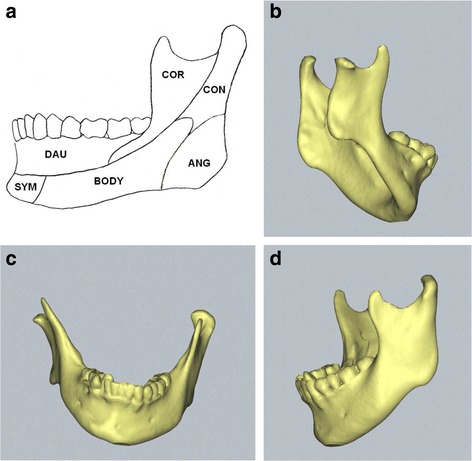
Background: The aim of this study was to present three-dimensional (3D) structural characteristics of the mandible in the hemifacial microsomia. The mandible has six distinct functional units, and its architecture is the sum of balanced growth of each functional unit and surrounding matrix.

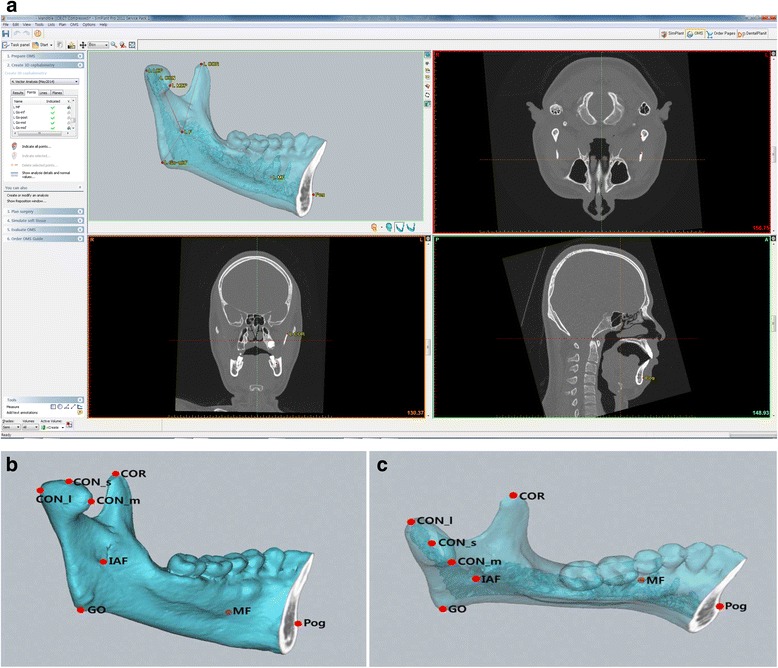
Methods: In order to characterize the mandibular 3D architecture of hemifacial microsomia, we analyzed the mandibular functional units of four hemifacial microsomia patients using the 3D reconstructed computed tomography (CT) images. And we compared the functional unit size between affected and non-affected side.

Results: The length of condyle and angle showed significant differences between affected and non-affected sides. However, the length of mandibular body showed insignificant differences. The size differences between affected and non-affected side were observed at the condyle, angle, and body in descending order.

Conclusions: This preliminary study suggests that the main etiopathogenic units are condyle and angle in the hemifacial microsomia mandible. Further investigation with the increased number of subjects will be helpful to establish treatment modality by etiopathogenic targeting of hemifacial microsomia.

Keywords: Computerized tomography; Functional unit; Hemifacial microsomia; Mandible; Three-dimensional.


Figures

Fig. 1
Fig. 1
Mandibular functional unit and three-dimensional computerized tomographic images of hemifacial microsomia mandible with Pruzansky’s type I deformity. a Mandibular functional units. b Affected side of hemifacial microsomia mandible. c Frontal view. d Non-affected side
Fig. 2
Fig. 2
Mandibular reference points for the mandibular functional unit analysis on the normal mandibular structure. a A screen capture image of the software used in this study (SimPlant, version 14.0, Materialise NV, Leuven, Belgium). b Lateral view of hemifacial microsomia mandible with reference points. c Occlusal view
Fig. 3
Fig. 3
Mandibular functional unit measurement with reference points on the normal mandibular structure. a Lateral view of hemifacial microsomia mandible. b Occlusal view

References

    1. Poswillo D. The pathogenesis of the first and second branchial arch syndrome. Oral Surg Oral Med Oral Pathol. 1973;35(3):302–328. doi: 10.1016/0030-4220(73)90070-4. - DOI - PubMed
    1. Carls FR, Sailer HF. Seven years clinical experience with mandibular distraction in children. J Craniomaxillofac Surg. 1998;26(4):197–208. doi: 10.1016/S1010-5182(98)80015-2. - DOI - PubMed
    1. Posnick JC. Surgical correction of mandibular hypoplasia in hemifacial microsomia: a personal perspective. J Oral Maxillofac Surg. 1998;56(5):639–650. doi: 10.1016/S0278-2391(98)90466-9. - DOI - PubMed
    1. Grabb WC. The first and second branchial arch syndrome. Plast Reconstr Surg. 1965;36(5):485–508. doi: 10.1097/00006534-196511000-00001. - DOI - PubMed
    1. Edgerton MT, Marsh JL. Surgical treatment of hemifacial microsomia. (First and second branchial arch syndrome) Plast Reconstr Surg. 1977;59(5):653–666. doi: 10.1097/00006534-197705000-00006. - DOI - PubMed

LinkOut - more resources