

Review
doi: 10.1155/2015/785206.
Epub 2015 Aug 27.
Added Value of Assessing Adnexal Masses with Advanced MRI Techniques
Affiliations
- PMID: 26413542
- PMCID: PMC4564594
- DOI: 10.1155/2015/785206
Item in Clipboard
Review
Added Value of Assessing Adnexal Masses with Advanced MRI Techniques
Biomed Res Int.
2015.
Abstract
This review will present the added value of perfusion and diffusion MR sequences to characterize adnexal masses. These two functional MR techniques are readily available in routine clinical practice. We will describe the acquisition parameters and a method of analysis to optimize their added value compared with conventional images. We will then propose a model of interpretation that combines the anatomical and morphological information from conventional MRI sequences with the functional information provided by perfusion and diffusion weighted sequences.
Figures
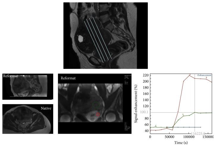
Perfusion MR acquisition. The sequence may be acquired in 2D plane in order to cover both external myometrium and the tumor or in axial 3D plane with a high quality of reformatting imaging in sagittal and coronal planes.
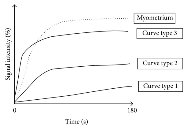
Time intensity curve.

Semiquantitative analysis. Different parameters published were EA (enhancement amplitude), THR (time of half rising), maximal slope (MS), SIrel (maximal relative enhancement), WIR (wash-in rate), and SImax.
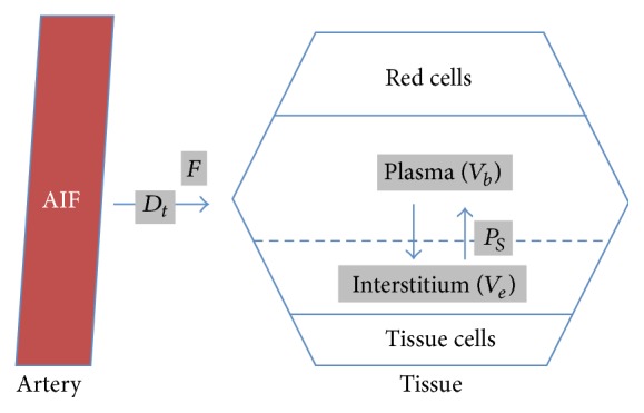
Pharmacokinetic model: Brix modified model with 4 quantitative parameters including tissue blood flow (F), blood volumetric fraction (V
b), the product of capillary wall permeability and surface area (P
S), interstitial volume (V
e), and the delay for the contrast media to reach tissue (D
t).
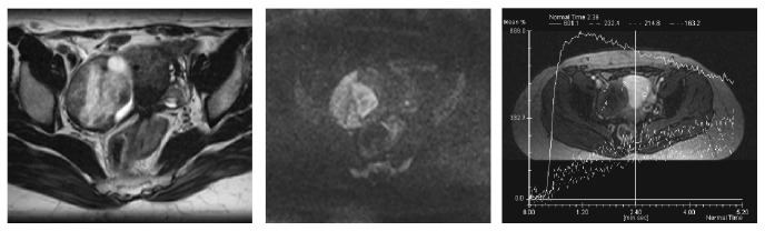
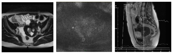
Ovarian fibroma right adnexal mixed cystic solid mass with an intermediate T2 weighted signal intensity in the solid component (A), a high DW signal (B), and a time intensity curve weak and progressive without any plateau (dotted line) in comparison with myometrial enhancement (continuous line).
T2 “dark though” effect—cystadenofibroma. The fibrous component of this tumor was highly cellular with an ADC value lower than 1 · 10−3 mm2/s. However, the tumor is not bright on DW image because of its low T2 signal (T2 dark through effect).
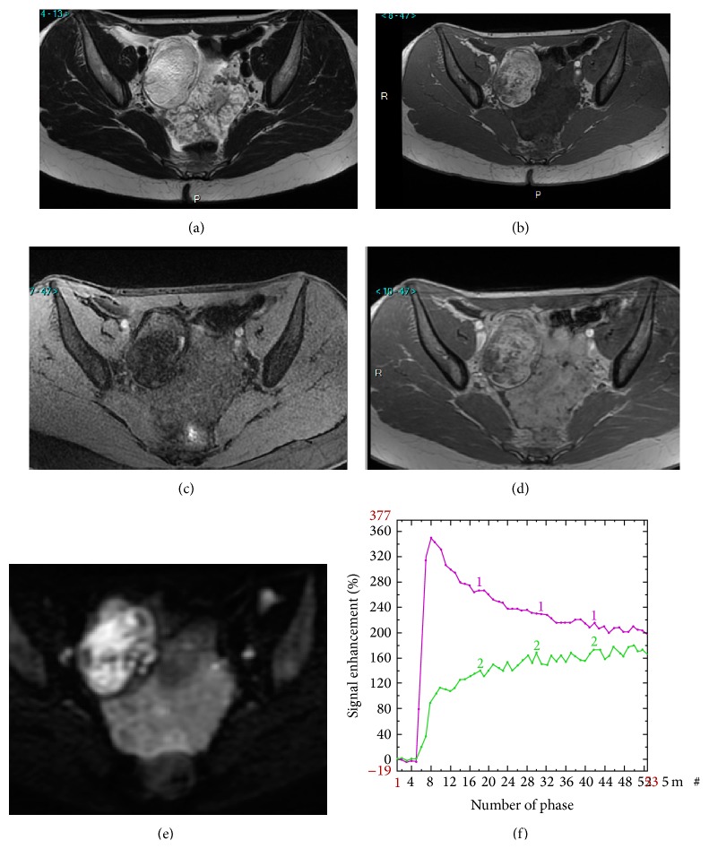
Added value of perfusion and diffusion weighted imaging. Right side: mature cystic teratoma (purely fatty mass: ADNEXMRscore 2). Left side: borderline serous cystadenoma (solid tissue which is bright on T2 and DW sequence and that enhances with a TIC type 2: ADNEXMRscore 4). T2 weighted sequence (a), T1 weighted sequence (b), T1 weighted sequence with fat saturation (c), T1 weighted sequence with gadolinium (d), DW sequence (e), and PW analysis comparing myometrial TIC (1) and tumoral TIC (2) (f).
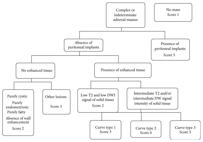
ADNEX MR scoring system (19).
References
-
- Emoto M., Iwasaki H., Mimura K., Kawarabayashi T., Kikuchi M. Differences in the angiogenesis of benign and malignant ovarian tumors, demonstrated by analyses of color Doppler ultrasound, immunohistochemistry, and microvessel density. Cancer. 1997;80(5):899–907. doi: 10.1002/(sici)1097-0142(19970901)80:560;899::aid-cncr1162;3.3.co;2-j. - DOI - PubMed
-
- Schutter E. M., Kenemans P., Sohn C. Diagnostic value of pelvic examination, ultrasound, and serum CA 125 in postmenopausal women with a pelvic mass. An international multicenter study. Cancer. 1994;74(4):1398–1406. - PubMed
-
- Vuento M. H., Pirhonen J. P., Makinen J. I., Laippala P. J., Gronroos M., Salmi T. A. Evaluation of ovarian findings in asymptomatic postmenopausal women with color doppler ultrasound. Cancer. 1995;76(7):1214–1218. doi: 10.1002/1097-0142(19951001)76:7x0003C;1214::aid-cncr2820760718x003E;3.0.co;2-5. - DOI - PubMed
Publication types
MeSH terms
Grants and funding
LinkOut - more resources
Full Text Sources
Other Literature Sources
Medical
