. 2015 Oct 27;6(33):35004-22.
doi: 10.18632/oncotarget.5253.

Valproic acid enhances the efficacy of radiation therapy by protecting normal hippocampal neurons and sensitizing malignant glioblastoma cells

Affiliations

Valproic acid enhances the efficacy of radiation therapy by protecting normal hippocampal neurons and sensitizing malignant glioblastoma cells

Dinesh Thotala et al. Oncotarget. .

Abstract

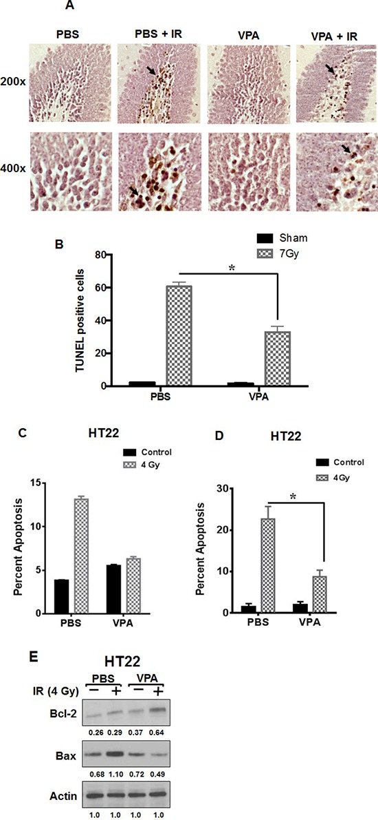
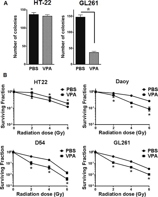
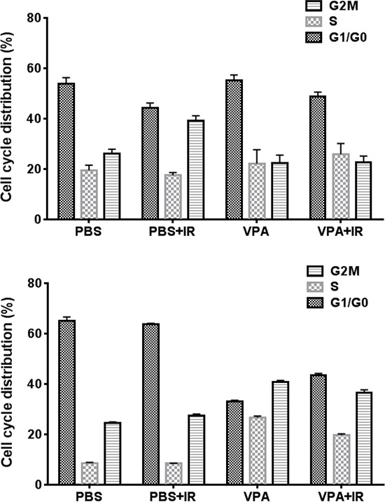
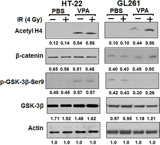
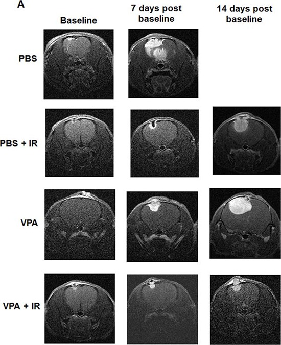
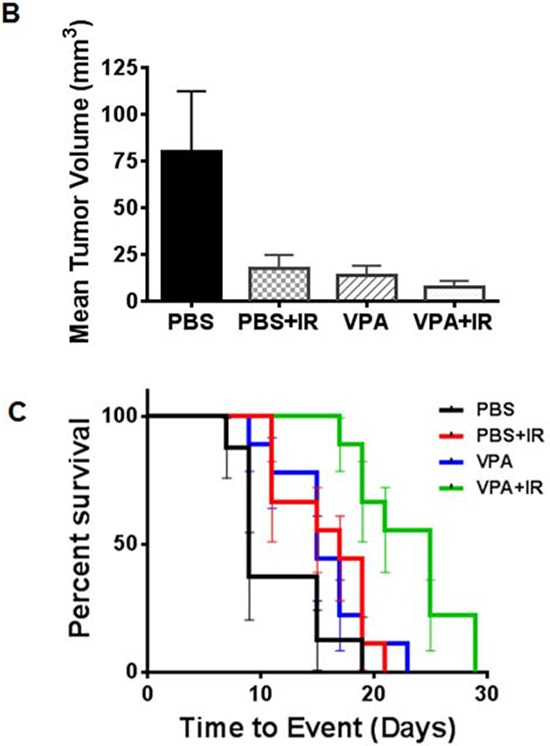
Neurocognitive deficits are serious sequelae that follow cranial irradiation used to treat patients with medulloblastoma and other brain neoplasms. Cranial irradiation causes apoptosis in the subgranular zone of the hippocampus leading to cognitive deficits. Valproic acid (VPA) treatment protected hippocampal neurons from radiation-induced damage in both cell culture and animal models. Radioprotection was observed in VPA-treated neuronal cells compared to cells treated with radiation alone. This protection is specific to normal neuronal cells and did not extend to cancer cells. In fact, VPA acted as a radiosensitizer in brain cancer cells. VPA treatment induced cell cycle arrest in cancer cells but not in normal neuronal cells. The level of anti-apoptotic protein Bcl-2 was increased and the pro-apoptotic protein Bax was reduced in VPA treated normal cells. VPA inhibited the activities of histone deacetylase (HDAC) and glycogen synthase kinase-3β (GSK3β), the latter of which is only inhibited in normal cells. The combination of VPA and radiation was most effective in inhibiting tumor growth in heterotopic brain tumor models. An intracranial orthotopic glioma tumor model was used to evaluate tumor growth by using dynamic contrast-enhanced magnetic resonance (DCE MRI) and mouse survival following treatment with VPA and radiation. VPA, in combination with radiation, significantly delayed tumor growth and improved mouse survival. Overall, VPA protects normal hippocampal neurons and not cancer cells from radiation-induced cytotoxicity both in vitro and in vivo. VPA treatment has the potential for attenuating neurocognitive deficits associated with cranial irradiation while enhancing the efficiency of glioma radiotherapy.

Keywords: cancer therapy; histone deacetylase (HDAC); neuroprotection; radioprotection; valproic acid (VPA).


Conflict of interest statement

CONFLICTS OF INTEREST

All authors declare no conflicts of interest.

Figures

Figure 1
Figure 1. VPA treatment protects hippocampal neurons from radiation-induced apoptosis in vivo and modulates the expression of apoptotic signaling proteins in vitro
A. One-week-old C57BL/6 mice pups were treated with daily i.p. injections of VPA (300 mg/kg) or PBS for 7 days. One hour after the last VPA treatment, the pups heads were irradiated with 7 Gy while the rest of the body was shielded with lead. Twenty-four hours later, the animals were sacrificed and brains were fixed and coronally sectioned. Sections that contained hippocampus were stained with TUNEL. Shown are representative photographs of mouse hippocampus. The arrows indicate examples of TUNEL positive cells (TPC). B. Eight HPF at 200x magnification were evaluated and TPC were counted for each experimental group. Shown is the average number of TPC per HPF for each radiation dose group (SD of three independent experiments, *P < 0.05). C. HT22 cells were treated with PBS or 0.6 mM VPA for 7 days prior to irradiation with 4 Gy. 24 h after irradiation, cells were stained with Annexin V-APC/propidium iodide and analyzed by flow cytometry; *P < 0.05 D. Cells were fixed and stained with DAPI, and apoptotic cells were counted in eight randomly selected HPF at 200X magnification. Shown are bar graphs of the average percent of apoptotic cells for each treatment with SD from three experiments; *P < 0.05. E. HT22 cells were treated with PBS or 0.6 mM VPA for 7 days prior to irradiation with 4 Gy. Whole cell extracts were immunobloted to determine the levels of Bax and Bcl-2. Actin was used to normalize the protein loading in each lane. Densitometry values representing the ratio of the various proteins normalized actin is indicated below each immunoblot.
Figure 2
Figure 2. VPA acts as a radioprotector in normal cells and radiosensitizer in cancer cells
A. Equal numbers of HT22 or GL261 cells were plated for colony formation assay after treating with PBS or 0.6 mM VPA for 7 days. Plates were stained with 1% methylene blue after 10 days and colonies were counted. Shown are bar graphs depicting the number of colonies for each treatment with SD from three experiments; *P < 0.05. B. HT22, Daoy, D54 and GL261 cell were treated with PBS (●) or 0.6 mM Valproic acid (■) for 7 days followed by irradiation with 0, 2, 4, 6 or 8 Gy and plated for clonogenic survival assay. Shown are the surviving fractions and the SD from three experiments; *P < 0.05. C. Equal numbers of HT22, Daoy, D54 and GL261 cells were plated in 96-well plates after treatment with PBS or 0.6 mM VPA for 7 days and then irradiated with 4Gy. After 96 h, the cell viability was determined using a colorimetric cell proliferation assay. Shown are the absorbances at 490 nm; *P < 0.05.
Figure 3
Figure 3. VPA treatment induces G2/M accumulation in cancer cells
HT22 cells and GL261 cells were treated with PBS or 0.6 mM VPA for 7 days prior to irradiation with 4 Gy. 24 h after irradiation, cells were collected, fixed and stained with PI and cell cycle distributions were determined by flow cytometry. Shown are the bar graphs of the average change in the fraction of G1/G0, S and G2/M phase cells after each treatment.
Figure 4
Figure 4. VPA inhibits both HDAC and GSK3β in normal cells, but only HDAC in cancer cells
HT22 and GL261 cells were treated with 0.6 mM VPA for 7 days prior to irradiation with 4 Gy. Total cellular proteins were immunobloted using antibodies against β-catenin, p-GSK3β (Ser9), GSK3β and Acetyl H4 to determine their levels. A representative (from 3 repeats) immunoblot is shown.
Figure 5
Figure 5. VPA delays tumor growth in irradiated mouse tumor models
GL261 or D54 cells were implanted into the right flank of nude mice. Once the tumors were palpable, they were irradiated with 2 Gy for 5 consecutive days for a total of 10 Gy. Mice were treated with VPA (300 mg/Kg) or PBS 60 min prior to irradiation. Tumor volumes were measured using digital calipers. Shown are the mean tumor volumes with SD from each treatment group of eight mice A & D, number of days to reach a tumor volume of 0.6 cm3 B & E, *P < 0.05, mean tumor volume following various treatments on day 15 C & F, *P < 0.05.
Figure 6
Figure 6. VPA in combination with irradiation improves survival by inhibiting tumor growth
Nude mice were implanted intracranially with GL261 cells stably expressing luciferase. After 10 days, mice were imaged with bioluminescence imaging and then serpentine sorted into four groups of nine each. Tumors were irradiated with 2 Gy or sham for five consecutive days shielding of the non-brain areas of the body during irradiation. Mice were treated with VPA (300 mg/Kg) or PBS 60 min prior to irradiation. A. Representative, contrast-enhanced, T1-weighted images of mouse brain from each of the four groups (top to bottom: PBS, PBS + IR, VPA, VPA + IR) at baseline (left) and 7 days (middle) and 14 days (right) post treatment. B. Average, MRI-derived tumor volumes measured at seven days post-treatment. C. Kaplan-Meyer survival curves following various treatments of mice bearing intracranial tumors.

References

    1. Liauw SL, Connell PP, Weichselbaum RR. New paradigms and future challenges in radiation oncology: an update of biological targets and technology. Science translational medicine. 2013;5:173-172. - PMC - PubMed
    1. Morris ZS, Harari PM. Interaction of radiation therapy with molecular targeted agents. J Clin Oncol. 2014;32:2886–2893. - PMC - PubMed
    1. Meadows AT, Gordon J, Massari DJ, Littman P, Fergusson J, Moss K. Declines in IQ scores and cognitive dysfunctions in children with acute lymphocytic leukaemia treated with cranial irradiation. Lancet. 1981;2:1015–1018. - PubMed
    1. Jannoun L, Bloom HJ. Long-term psychological effects in children treated for intracranial tumors. Int J Radiat Oncol Biol Phys. 1990;18:747–753. - PubMed
    1. Welzel G, Fleckenstein K, Mai SK, Hermann B, Kraus-Tiefenbacher U, Wenz F. Acute neurocognitive impairment during cranial radiation therapy in patients with intracranial tumors. Strahlenther Onkol. 2008;184:647–654. - PubMed

Publication types

MeSH terms