. 2016 Jan;64(1):139-54.
doi: 10.1002/glia.22921. Epub 2015 Sep 28.

The speed of swelling kinetics modulates cell volume regulation and calcium signaling in astrocytes: A different point of view on the role of aquaporins

Affiliations

The speed of swelling kinetics modulates cell volume regulation and calcium signaling in astrocytes: A different point of view on the role of aquaporins

Maria Grazia Mola et al. Glia. 2016 Jan.

Abstract

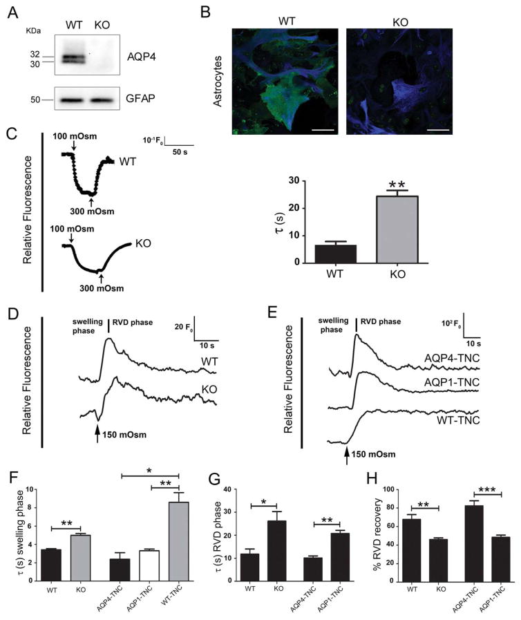
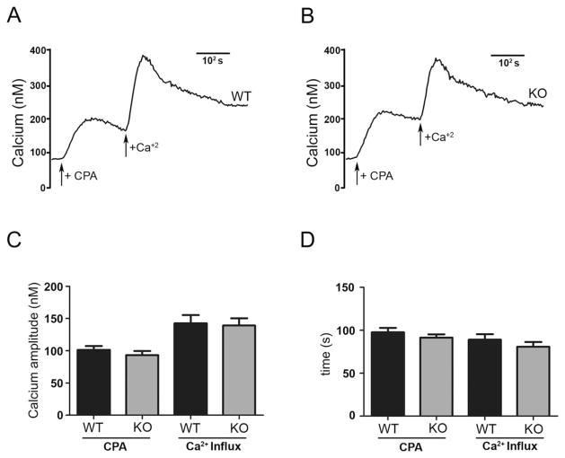
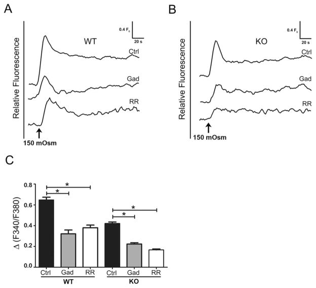
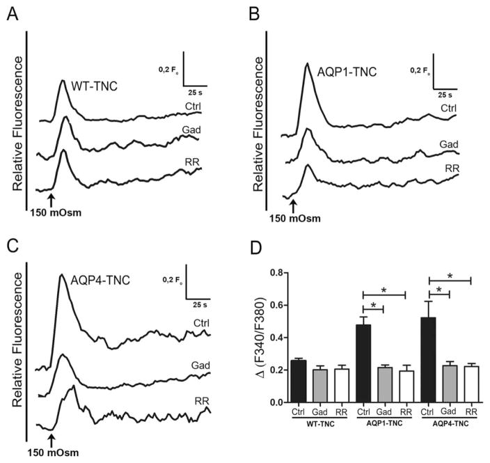
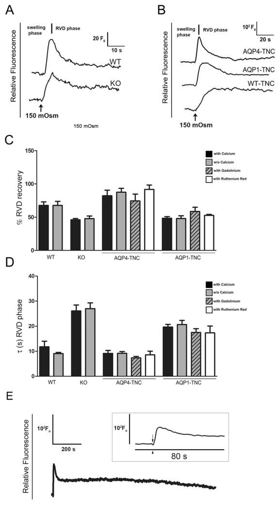
Regulatory volume decrease (RVD) is a process by which cells restore their original volume in response to swelling. In this study, we have focused on the role played by two different Aquaporins (AQPs), Aquaporin-4 (AQP4), and Aquaporin-1 (AQP1), in triggering RVD and in mediating calcium signaling in astrocytes under hypotonic stimulus. Using biophysical techniques to measure water flux through the plasma membrane of wild-type (WT) and AQP4 knockout (KO) astrocytes and of an astrocyte cell line (DI TNC1) transfected with AQP4 or AQP1, we here show that AQP-mediated fast swelling kinetics play a key role in triggering and accelerating RVD. Using calcium imaging, we show that AQP-mediated fast swelling kinetics also significantly increases the amplitude of calcium transients inhibited by Gadolinium and Ruthenium Red, two inhibitors of the transient receptor potential vanilloid 4 (TRPV4) channels, and prevented by removing extracellular calcium. Finally, inhibition of TRPV4 or removal of extracellular calcium does not affect RVD. All together our study provides evidence that (1) AQP influenced swelling kinetics is the main trigger for RVD and in mediating calcium signaling after hypotonic stimulus together with TRPV4, and (2) calcium influx from the extracellular space and/or TRPV4 are not essential for RVD to occur in astrocytes.

Keywords: AQP1; AQP4; RVD; cell swelling; hypotonic stimulus.


Figures

FIGURE 1
FIGURE 1
Water transport and RVD in primary cultured astrocytes endogenously expressing AQP4 and in DI TNC1 transfected with AQP4 or AQP1. (A) Western blots showing AQP4 and GFAP expression in WT and AQP4 KO (KO) astrocyte primary cultures. Note the expression of both M1 andM23-AQP4 isoforms in WT and their absence in AQP4 KO astrocytes. GFAP was used as the internal standard for protein loading. (B) Merged confocal microscopy images of WT and AQP4 KO (KO) astrocytes immunostained for AQP4 (green) and GFAP (blue). Note the absence of AQP4 staining in KO and the typical punctate AQP4 staining in WT (Scale bar=50 μm). (C) (Left) Time course of water transport recorded from Calcein-AM loaded WT and AQP4 KO (KO) astrocytes using TIRF microscopy showing changes in fluorescence induced by 200 mOsm L−1 hypotonic gradient. The arrows indicate the addition of a hypotonic solution (100 mOsm L−1) and isotonic solution (300 mOsm L−1). (Right) Histogram of the mean±SE values of time constants obtained by fitting an exponential curve to the cell swelling phase of cells after exposure to 100 mOsm L−1 solution (**P< 0.005; n=4). (D,E) Functional analysis for plasma membrane water transport performed by using Flex Station with the Calcein-AM quenching assay, showing the typical swelling kinetics (swelling phase) followed by the RVD kinetics (RVD phase) ofWT and AQP4 KO (KO) astrocytes (D) and DI TNC1 transfected with AQP4 (AQP4-TNC) or AQP1 (AQP1-TNC) and WT (WT-TNC) (E). (F) Histogram showing the mean±SE values of the time constants obtained for the “swelling phase” of astrocytes and DI TNC1 cells exposed to 150 mOsm L−1 hypotonic solution, obtained using the Calcein-AM quenching assay. (G, H) Histograms showing the mean±SE values of the time constant (G) and of the extent of recovery (in percent, H) of the RVD phase of the indicated cells. Note that AQP expression promotes faster RVD kinetics and higher efficiency of RVD (F–H: ***P < 0.0005; **P< 0.005; *P< 0.05; n=6 forWT; n=8 for KO; n=5 for AQP4-TNC; n=8 forAQP1-TNC; n=9 for WT-TNC).
FIGURE 2
FIGURE 2
Hypotonic stimulus induced intracellular calcium rise in WT and AQP4 KO astrocytes. (A,B) Time courses of intracellular calcium concentration changes recorded from WT (A) and AQP4 KO (B) astrocytes exposed to calcium-containing hypotonic solution. Note the delayed and reduced amplitudes of calcium responses in AQP4 KO astrocytes compared to those of WT cells. (C,D) Time course of intracellular calcium concentration changes recorded from WT (C) and AQP4 KO (D) astrocytes exposed to calcium-free hypotonic solution. Note that for both WT and AQP4 KO astrocytes, calcium amplitudes are dramatically reduced compared to those recorded in calcium-containing solution (A,B). (E,F) Histograms showing the mean±SE values of calcium amplitude (E) and time to peak (F) recorded from WT and AQP4 KO astrocytes exposed to hypotonic calcium containing (with Ca2+) and calcium-free (w/o Ca2+) solution (*P < 0.05, n=6).
FIGURE 3
FIGURE 3
Intracellular calcium content and calcium release activated channels are not altered in AQP4 KO astrocytes. (A,B) Kinetics of cytosolic calcium changes induced by CPA (+CPA) and following addition of calcium containing external solution (+Ca2+) in WT (A) and AQP4 KO (B) astrocytes. (C,D) Histograms showing the mean±SE values of the amplitudes (C) and time to peak (D) of calcium responses, after store depletion by CPA and addition of extracellular calcium (n=5).
FIGURE 4
FIGURE 4
Gadolinium and Ruthenium Red partially block calcium increase induced by hypotonicity-induced astrocyte swelling. Superimposed kinetics of calcium responses recorded from untreated (Ctrl) and Gadolinium (Gad) or Ruthenium Red (RR) treated WT (A) and AQP4 KO (B) astrocytes exposed to hypotonic stimulus. (C) Histograms showing the mean±SE values of the calcium amplitudes measured under conditions shown in A and B. Note that in both WT and AQP4 KO astrocytes Gadolinium and Ruthenium Red significantly reduced the amplitude of intracellular calcium levels induced by hypotonic stimulus (*P < 0.05, n=6).
FIGURE 5
FIGURE 5
Intracellular calcium response under hypothonic stimulus in WT and AQP1 or AQP4 transfected DI TNC1 treated with Gadolinium or Ruthenium Red. Superimposed kinetics of calcium concentration after hypotonic stimulus of WT (A), AQP1 transfected (B) and AQP4 transfected (C) DI TNC1, untreated (Ctrl) or treated with Gadolinium (Gad) or Ruthenium Red (RR). (D) Histograms showing the mean values±SE of calcium amplitude of the conditions shown in A, B and C. Note that when added separately both Gadolinium and Ruthenium Red significantly affect the amplitude of intracellular calcium increase in both AQP4 and AQP1 transfected cells (*P < 0.05, n=5).
FIGURE 6
FIGURE 6
RVD in WT, AQP4 KO astrocytes and AQP expressing DI TNC1 is not dependent on extracellular calcium and TRPV4. (A,B) Kinetics of RVD recorded from astrocyte primary cultures (A) and DI TNC1 (B) exposed to calcium-free hypotonic solution. (C,D) Histograms showing the mean±SE values of the extent of recovery (in percent) (C) and time constant (D) of RVD of the indicated cells, in the presence (black bars) and in the absence (grey bars) of external calcium, and in cells treated with Gadolinium (striped bars) or Ruthenium Red (white bars) in the presence of external calcium. In each cell type no significant differences were detected between the conditions analyzed. (E) Longer RVD kinetics recorded from astrocyte primary cultures for 1,200 s. The curve in the inset shows that, immediately after the 150 mOsm L−1 hypotonic stimulus (arrow) given at 17 s (arrowhead), the cells undergo a fast RVD which takes <40 s. Recording data for 1,200 s demonstrate that there is a further volume decrease, although without a total recovery.
FIGURE 7
FIGURE 7
Schematic representation of the mechanism proposed to describe the different effect of slow and fast swelling kinetics on calcium response and RVD. The absence (A) or presence (B) of AQP water channels in the plasma membrane affects the cell swelling kinetics which is slow (A) and fast (B), respectively. The swelling kinetics modulates the level of plasma membrane stretch, here symbolized by arrows parallel to the plasma membrane. (A) Slow swelling. The swelling in the absence of AQP water channels can be very slow (see WT-TNC), depending on the composition of the lipid bilayer through which the water influx occurs by simple diffusion (1), and produces a weak plasma membrane stretch (thin arrows). This weak stretch is sufficient for the opening of TRPV4 channels (2), with a consequent calcium influx, but not sufficient to open SAC (Stretch Activated Channels), which are responsible for the cell volume regulation, and therefore no RVD occurs. (B) Fast swelling. The swelling in the presence of AQP water channels is always fast (see AQP-TNC or WT astrocyte primary cultures). In this case, AQP mediated water influx (1) is characterized by fast swelling kinetics producing strong plasma membrane stretch (thick arrows). The membrane stretch in this case is strong enough (a) to induce a major opening of TRPV4 (2), with consequent increased calcium influx, compared to the situation described in A, and (b) to open calcium independent SAC (2) responsible for the cell volume regulation through the efflux of ions/solute. This efflux of ions/solute is followed by a water efflux through AQP water channels (3) itself responsible for the RVD.

References

    1. Agre P, King LS, Yasui M, Guggino WB, Ottersen OP, Fujiyoshi Y, Engel A, Nielsen S. Aquaporin water channels—From atomic structure to clinical medicine. J Physiol. 2002;542:3–16. - PMC - PubMed
    1. Altamirano J, Brodwick MS, Alvarez-Leefmans FJ. Regulatory volume decrease and intracellular calcium in murine neuroblastoma cells studied with fluorescent probes. J Gen Physiol. 1998;112:145–160. - PMC - PubMed
    1. Amiry-Moghaddam M, Ottersen OP. The molecular basis of water transport in the brain. Nat Rev Neurosci. 2003;4:991–1001. Review. - PubMed
    1. Amiry-Moghaddam M, Xue R, Haug FM, Neely JD, Bhardwaj A, Agre P, Adams ME, Froehner SC, Mori S, Ottersen OP. Alpha-syntrophin deletion removes the perivascular but not endothelial pool of aquaporin-4 at the blood-brain barrier and delays the development of brain edema in an experimental model of acute hyponatremia. FASEB J. 2004;18:542–544. - PubMed
    1. Basco D, Blaauw B, Pisani F, Sparaneo A, Nicchia GP, Mola MG, Reggiani C, Svelto M, Frigeri A. AQP4-dependent water transport plays a functional role in exercise-induced skeletal muscle adaptations. PLoS One. 2013;8:e58712. doi: 10.1371/journal.pone.0058712. Erratum in: PLoS One 8(6). doi:10.1371/annotation/86fc2632-913c-490d-8b9b-e925b38baec5. - DOI - PMC - PubMed

Publication types

MeSH terms