Review
. 2015 Dec 9;27(46):7620-37.
doi: 10.1002/adma.201501810. Epub 2015 Sep 28.

Conducting Polymers for Neural Prosthetic and Neural Interface Applications

Affiliations
Review

Conducting Polymers for Neural Prosthetic and Neural Interface Applications

Rylie Green et al. Adv Mater. .

Abstract

Neural-interfacing devices are an artificial mechanism for restoring or supplementing the function of the nervous system, lost as a result of injury or disease. Conducting polymers (CPs) are gaining significant attention due to their capacity to meet the performance criteria of a number of neuronal therapies including recording and stimulating neural activity, the regeneration of neural tissue and the delivery of bioactive molecules for mediating device-tissue interactions. CPs form a flexible platform technology that enables the development of tailored materials for a range of neuronal diagnostic and treatment therapies. In this review, the application of CPs for neural prostheses and other neural interfacing devices is discussed, with a specific focus on neural recording, neural stimulation, neural regeneration, and therapeutic drug delivery.

Keywords: conducting polymers; neural interfaces; neural recording; neural stimulation.


Figures

Figure 1
Figure 1
Schematics showing cellular responses during early (A) and sustained (B) reactive responses observed following neural device insertion. The early response (A) is characterized by a large region containing reactive astrocytes and microglia around inserted devices. The sustained response (B) is characterized by a compact sheath of cells around insertion sites. Inserts depict potential cell–cell interactions and signaling path ways. Neurons (pink), astrocytes (red), monocyte derived cells including microglia (blue), and vasculature (purple) are depicted. (C), (D) GFAP immunohistochemistry (Marker for astrocytes) of tissues slices from brains and (E), (F) ED1 immunohistochemistry (marker for microglia) of tissues slices due to the implantation of neural electrodes.. GFAP-immunohistochemistry (red) and nuclear staining (green) of devices removed from brains at 1 day (G) and 1 (H), 6 (I), and 12 (J) weeks post insertion. All scale bars = 100 μm. Reproduced with permission.[17] Copyright 2003.
Figure 2
Figure 2
(A) Chemical structures of various conducting polymers PPy, PEDOT, PT, PANI. (B) Polymerization of PEDOT and doped with anion (dopant) A. (C) Oxidation and reduction behavior of conducting polymers are dependent on dopant size. Small anion A is able to migrate in and out of the PPy matrix to balance the charge across the backbone. Large anion B is immobilized within the PPy matrix and hence relies on smaller cation X. to migrate into the polymer from the surrounding electrolyte and maintain charge balance across the polymer. Reproduced with permission.[50] Copyright 2009.
Figure 3
Figure 3
(A) Schematic of ideal hybrid configuration and photo comparison of hybrid material created from using a bound dopant (B), compared to stratified composite produced from using a free dopant (C), both material samples are hydrated. (D) Charge storage capacity of hybrids calculated from CV performed versus Ag/AgCl at 120mV s−1 from −700 to 700mV over 850 cycles. (E) Elastic moduli (calculated from DMT model) under hydrated conditions, compared to Pt, homogeneous CPs and neural tissue. Reproduced with permission.[13] Copyright 2012. (F) Diameters of the PLGA fibers were distributed over the range 40–500 nm with the majority being between 100–200 nm. (G) Electropolymerized PEDOT nanotubes on the electrode site of an acute neural probe tip after removing the PLGA core fibers. (H) A section of (G) cut with a focused ion beam showing the silicon substrate layer and PEDOT nanoscale fiber coating. (I) Higher-magnification image of (H) showing the PEDOT nanotubes crossing each other. (J) A single PEDOT nanotube which was polymerized around a PLGA nanoscale fiber, followed by dissolution of the PLGA core fiber. This image shows the external texture at the surface of the nanotube. (K) Higher-magnification image of a single PEDOT nanotube demonstrating the textured morphology that has been directly replicated from the external surface of the electrospun PLGA fiber templates. The average wall thickness of PEDOT nanotubes varied from 50–100 nm, with the nanotube diameters ranging from 100 to 600 nm. Schematic illustration of the controlled release of dexamethasone: (L) dexamethasone-loaded electrospun PLGA, (M) hydrolytic degradation of PLGA fibers leading to release of the drug, and (N) electrochemical deposition of PEDOT around the dexamethasone-loaded electrospun PLGA fiber slows down the release of dexamethasone. (O) PEDOT nanotubes in a neutral electrical condition. (P, Q) External electrical stimulation controls the release of dexamethasone from the PEDOT nanotubes due to contraction or expansion of the PEDOT. By applying a positive voltage, electrons are injected into the chains and positive charges in the polymer chains are compensated. To maintain overall charge neutrality, counterions are expelled towards the solution and the nanotubes contract. This shrinkage causes the drugs to come out of the ends of tubes. (R) Cumulative mass release of dexamethasone from: PLGA nanoscale fibers (black squares), PEDOT-coated PLGA nanoscale fibers (red circles) without electrical stimulation, and PEDOT-coated PLGA nanoscale fibers with electrical stimulation of 1 V applied at the five specific times indicated by the circled data points (blue triangles). (S) UV absorption of dexamethasone- loaded PEDOT nanotubes after 16 h (black), 87 h (red), 160 h (blue), and 730 h (green). The UV spectra of dexamethasone have peaks at a wavelength of 237 nm. Data are shown with a ± standard deviation (n = 15 for each case). Reproduced with permission.[68] Copyright 2006.
Figure 4
Figure 4
(A) Procedure for hydrogel coating the neural electrodes. Optical images of reswelling of the hydrogel-coated electrodes in an agar (1%)-phantom matrix as a function of time. HG coatings in the agar matrix made of (B) PBS and (C). deionized water. (C) Plot of reswelling of hydrogel coatings in the PBS agar. The initial thickness of hydrogel coatings was 50 lm as determined by optical microscopy during the dipcoating process. (E) The average percentages of clearly detectable units as a function of the thickness of hydrogel-coated electrodes in the auditory cortex with a 200 ms noise burst. The HG thickness indicates the initial thickness of hydrogel coatings before drying. The inset shows a representative recorded signal with a SNR of 5.0. (*) Significance between control (bare electrode) and hydrogel-coated electrodes. Reproduced with permission.[5] Copyright 2010
Figure 5
Figure 5
Laser micromachined arrays used in studies: (A) nine-electrode array used for in vitro studies with 7 PEDOT/pTS-coated sites (dark) and two bare Pt sites (light); (B) 24-electrode array in hexagonal (hex) configuration used for in vivo studies with 12 bare Pt (light electrode sites) and 12 PEDOT/pTS-coated electrodes (dark electrodes). SEM of (C) bare Pt electrodes, (D) coated with PEDOT/pTS at 200Å~ magnification and (E) PEDOT/pTS at 1000Å~ magnification. (F) Average maximum voltage drop across electrodes, measured in PBS with small biphasic pulse (100 μA, 100 μs) versus large, low impedance Pt counter. Each data point represents the mean voltage drop across 50 individual electrode sites and error bars represent one standard deviation (n = 50). (G) Average voltage drop across PEDOT electrodes compared to Pt electrodes during a 36 h acute study. Each data point represents the mean voltage of 12 electrodes with error bars showing one standard deviation (n = 12). Reproduced with permission.[7] Copyright 2013
Figure 6
Figure 6
PC-12 cell differentiation on PPy without (A) and with (B) application of an electric potential. PC-12 cells were grown on PPy for 24 h in the presence of NGF, then exposed to electrical stimulation (100 mV) across the polymer film, S (B). Images were acquired 24 h after stimulation. Cells grown for 48 h but not subjected to electrical stimulation, NS, are shown for comparison (A). Bar= 100 μm. Neurite length histograms. Shown are histograms of neurite lengths for cells on PPy with (S, stimulated) (C) and without (NS, unstimulated) (D) potential applied through PPy, on PPy with potential applied through the solution (E), and on tissue culture polystyrene (TCPS) (F). Reproduced with permission.[45] Copyright 1999. Characterization of the multilayered PEDOT:PSS conducting nanoparticles and their assembly as linear conduits. (G, H) SEM micrographs of the nanoparticle patterns at a magnification of 25kX (G) and 11kX (H) indicating the formation of tightly packed and highly ordered nanoparticle arrays. (I) Confocal fluorescence image of the RhB functionalized PEDOT:PSS nanoparticle arrays at 20X magnification. (J) High magnification (12kX magnification) SEM images demonstrating specific and preferential interactions of neurites (white arrows) with the PEDOT:PSS linear conduits (red arrows). (K) High-magnification SEM image (magnification 8kX) indicating the formation of extensive dendritic networks (white arrows) guided by the PEDOT:PSS arrays. Inset: The corresponding low-magnification image of the area (yellow box) analyzed (magnification 3kX). (L) Significant increase in the average cell area is observed 72 h after exogenous electrical stimulation on the nanoparticle platform in comparison to unstimulated and non-patterned controls. (M) Corresponding decrease in PC12 cell proliferation observed 72 h after exogenous electrical stimulation on the nanoparticle platform in comparison to unstimulated and non-patterned controls. Reproduced with permission.[136] Copyright 2015.
Figure 7
Figure 7
Scanning electron micrographs of electrospun PLGA nanofibers, (A) and (B), and their PPy-coated fibers, (C) and (D). (A) Randomly oriented nanofibers (RF), (B) aligned nanofibers (AF), (C) PPy-coated randomly oriented fibers (PPy–RF), and (D) PPy-coated aligned fibers (PPy–AF). SEM images of PC12 cells cultured on (E) PPy–RF and (F) PPy–AF for 2 days. (G) Median neurite lengths PC12 cells when unstimulated and when electrically stimulated (10 mV/cm) on random (PPy–RF) and aligned (PPy–AF) PPy–PLGA fibers. At least 300 neurites were analyzed from four substrates for each condition. Reproduced with permission.[137] Copyright 2009. SEM at 15,000_ magnification of NGF entrapped in PEDOT films compared with control films produced without NGF modification of the electrolyte (H) PEDOT/pTS, (I) PEDOT/DEDEDYFQRYLI, (J) PEDOT/DCDPGYIGSR, (K) PEDOT/pTS/NGF, (L) PEDOT/DEDEDYFQRYLI/NGF, (M) PEDOT/DCDPGYIGSR/NGF. (N) Electrochemical activity loss for NGF-loaded PEDOT films over 400 cycles of redox. Loss of electroactivity is plotted as a percentage of the original activity measured at cycle 1, normalising the polymers to a common baseline. The percentage activity remaining after 400 cycles is labeled. Error bars represent the standard error of the mean (n = 3). (O) Effect of NGF loading on ASTM hardness of PEDOT-based polymers. Gouge hardness is representative of a minor disruption to the polymer surface and scratch hardness depicts the level at which no polymer was removed from the Pt substrate. Error bars represent the standard error of the mean (n = 6). (P) Neurite outgrowth of PC12 cells on laminin peptide-doped PEDOT with NGF incorporation at 96 h post-plating. Neurite outgrowth is represented as the neurite length per adhered cell, calculated by normalising the total neurite length to the cell density. The standard error of the mean is given (n = 3). *No significant difference. Reproduced with permission.[49] Copyright 2010
Figure 8
Figure 8
Schematic illustration of fabrication process and optical micrographs of FPEDOTA and PPEDOTA conduits. FPEDOTA: A) PS template fiber (gray color in cross section (a)). B) Sputter coating the PS fiber with thin layer of AuPd (yellow color in cross section (b)). C) Dipping into the agarose solution (blue color in cross section (c)). D) Formation of agarose layer (blue color in cross section (d)). E) Electropolymerization of PEDOT on the surface of the AuPd-coated PS fibers (black color in cross section (e)). F) Dissolving the PS fiber (white color in cross section (f)). G) Electropolymerization of PEDOT on the surface of AuPd within the inner lumen (black color in cross section (g)). PPEDOTA: H) PS template fiber (gray color in cross section (h,i)). I) Wrapping of masking tape in spiral fashion around the PS fiber. J) Sputter coating the PS fiber with thin layer of AuPd (yellow color in cross section (j,k)). K) Removal of masking tape. L) Dipping into the agarose solution (blue color in cross section (l)). M) Formation of agarose layer (blue color in cross section (m). N) Electropolymerization of PEDOT on the surface of the AuPd-coated PS fibers (black color in cross section (n)). O) Dissolving the PS fiber (white color in cross section (o)). P) Electropolymerization of PEDOT on the surface of AuPd within the inner lumen (black color in cross section (p)). Q–R) Optical micrographs of FPEDOTA conduits with different magnifications. Red arrows show growth of PEDOT within the agarose hydrogel (R). S–T) Optical micrographs of PPEDOTA conduits with different magnify cations before electrodeposition PEDOT on the surface of AuPd within the inner lumen. Optical micrographs of implanted conduits, and EDL maximal specific muscle force. (U,V) Optical images of implanted FPEDOTA conduits in 10 mm peroneal nerve gap in rats. (W,X) Optical images of implanted PA conduits in 10 mm peroneal nerve gap in rats. (Y) Bar graph maximal specific muscle force. Column height represents the mean while error bars reflect the standard deviation of the mean (n = 5). The arrows show the location of stents. Optical micrographs of peripheral nerves at midgraft. (1, 2) Autograft. (3, 4) PA. (5, 6) FPEDOTA. (7, 8) PPEDOTA. Scale bars are 50 μm. Reproduced with permission.[110] Copyright 2012

References

    1. Besterman R Creese. Waller--pioneer of electrocardiography. Br Heart J. 1979;42:61–64. - PMC - PubMed
    1. Wark HAC, Sharma R, Mathews KS, Fernandez E, Yoo J, Christensen B, Tresco P, Rieth L, Solzbacher F, Normann RA, Tathireddy P. A new high-density (25 electrodes/mm 2 ) penetrating microelectrode array for recording and stimulating sub-millimeter neuroanatomical structures. Journal of Neural Engineering. 2013;10:045003. - PubMed
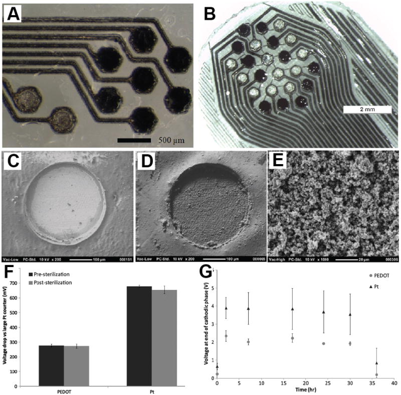
    1. Venkatraman S, Hendricks J, King ZA, Sereno AJ, Richardson-Burns S, Martin D, Carmena JM. In Vitro and In Vivo Evaluation of PEDOT Microelectrodes for Neural Stimulation and Recording. Neural Systems and Rehabilitation Engineering, IEEE Transactions on. 2011;19:307–316. - PubMed
    1. Ludwig KA, Uram JD, Yang J, Martin DC. Chronic neural recordings using silicon microelectrode arrays electrochemically deposited with poly(3,4-ethylenedioxythiophene) (PEDOT) film. J Neural Eng. 2006;3:59–70. - PubMed
    1. Kim D-H, Wiler JA, Anderson DJ, Kipke DR, Martin DC. Conducting polymers on hydrogel-coated neural electrode provide sensitive neural recordings in auditory cortex. Acta Biomaterialia. 2010;6:57–62. - PubMed

Publication types