. 2015 Nov;17(11):1497-1503.
doi: 10.1038/ncb3248. Epub 2015 Sep 28.

Lateral adhesion drives reintegration of misplaced cells into epithelial monolayers

Affiliations

Lateral adhesion drives reintegration of misplaced cells into epithelial monolayers

Dan T Bergstralh et al. Nat Cell Biol. 2015 Nov.

Abstract

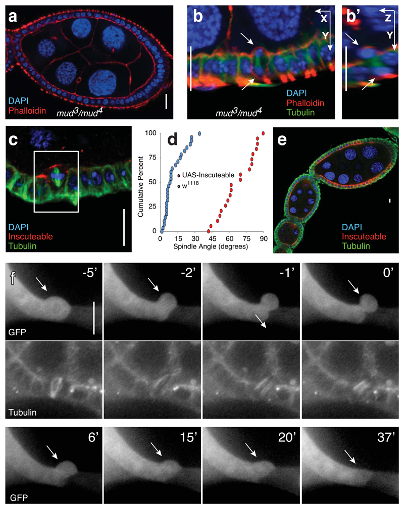
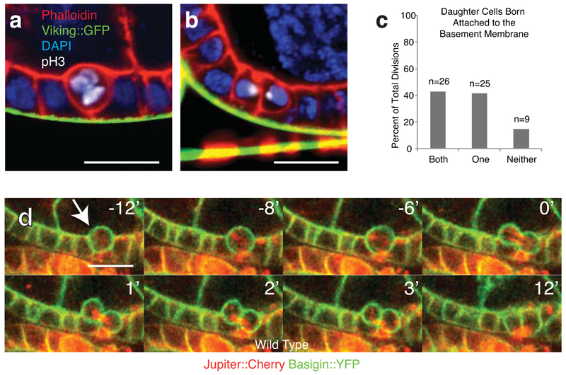
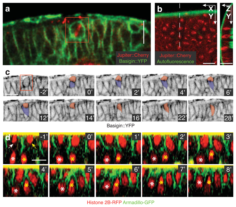
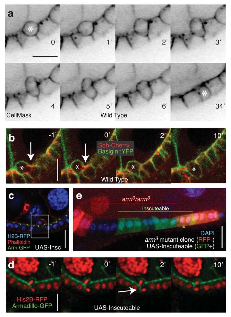
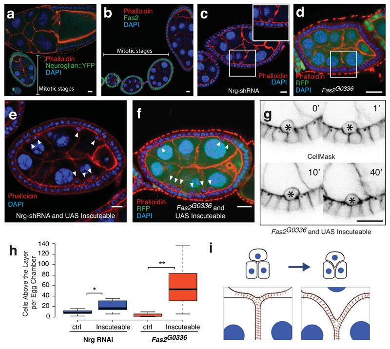
Cells in simple epithelia orient their mitotic spindles in the plane of the epithelium so that both daughter cells are born within the epithelial sheet. This is assumed to be important to maintain epithelial integrity and prevent hyperplasia, because misaligned divisions give rise to cells outside the epithelium. Here we test this assumption in three types of Drosophila epithelium; the cuboidal follicle epithelium, the columnar early embryonic ectoderm, and the pseudostratified neuroepithelium. Ectopic expression of Inscuteable in these tissues reorients mitotic spindles, resulting in one daughter cell being born outside the epithelial layer. Live imaging reveals that these misplaced cells reintegrate into the tissue. Reducing the levels of the lateral homophilic adhesion molecules Neuroglian or Fasciclin 2 disrupts reintegration, giving rise to extra-epithelial cells, whereas disruption of adherens junctions has no effect. Thus, the reinsertion of misplaced cells seems to be driven by lateral adhesion, which pulls cells born outside the epithelial layer back into it. Our findings reveal a robust mechanism that protects epithelia against the consequences of misoriented divisions.


Conflict of interest statement

Competing Financial Interest

The authors declare no competing financial interests.

Figures

Fig. 1
Fig. 1. Spindle misorientation does not disrupt the organisation of the follicle epithelium
A) The organisation of the follicle cells into an epithelial monolayer is normal in mud3/mud4 transheterozygotes. Disorganization was not observed in any of 42 ovarioles imaged. B) Incorrectly oriented divisions are observed in mud mutant egg chambers. Five such complete reorientations were observed. These are exceptional cases, given that mud mutation randomizes, rather that perpendicularizes, divisions. C) Ectopically expressed Inscuteable localizes apically and causes spindle reorientation in follicle cells. This image is one of the 20 divisions quantified in Figure 1D. D) A cumulative data plot of spindle angles in wild type (n = 29 spindles assessed from 29 different egg chambers) and UAS-Inscuteable expressing (n = 20 spindles assessed from 20 different egg chambers) follicle cells shows the extent of spindle misorientation caused by Inscuteable expression. p = 1.047x10⁻¹⁰ as determined by the Kolmogorov-Smirnof test. E) Inscuteable expression does not disrupt the organisation of the follicular epithelium. This image is representative of 103 ovarioles imaged. F) The apical product of a misoriented division reintegrates into the monolayer. Clonal expression of Actin-Gal4 was used to drive both UAS-Inscuteable and UAS-GFP, which illuminates the relevant cell (arrow) and its neighbors to the left. Tubulin-RFP marks the misoriented spindle at time -5’. As in subsequent timelapses, the 0’ timepoint represents abscission. This is one of seven complete reintegrations imaged. Scale bars in this figure represent 10μM.
Fig. 2
Fig. 2. Reintegration is a feature of wild type cell division in the follicle epithelium.
A and B) During division cells usually move apically relative to the epithelial layer, often appearing to detach detaching from the basement membrane (Viking-GFP). One prometaphase cell is shown in (A) and two recently born sister cells in (B). These fixed-tissue images are representative of the live divisions quantified in (C). C) Cells are frequently born without an obvious connection to the basement membrane. Divisions were imaged live to a depth of at least 10 microns to confirm the position of each cell in the tissue. n = number of cell divisions. D) Reintegration occurs in wild type tissue. Egg chambers were imaged live using Jupiter:Cherry to mark the spindle and Basigin-YFP to mark the cell outline. Images taken from Supplementary Movie 2. Timepoints in this and all subsequent timecourses are numbered relative to abcission (at zero). This movie is one of the 60 divisions quantified in C. Scale bars in this figure represent 10μM.
Fig. 3
Fig. 3. Reintegration occurs in multiple epithelia
A) Inscuteable expression causes spindle misorientation in the optic lobe neuroepithelium. The two daughter cells are tracked in (C). This image is representative of the Inscuteable-induced spindle misorientation in this tissue quantified in Egger et al. B) Expression of Inscuteable reorients mitotic spindles in the early embryonic ectoderm. Spindles are perpendicular to the plane of the epithelium. These spindles are among the 34 quantified in Supplemental Figure 3B. C) Both products of a misoriented division are maintained in the neuroepithelial layer. Pseudo-colouring indicates the two division products. By the 12’ minute timepoint, the basal daughter has reattached to the apical surface. After this point it moves orthogonally to the plane of focus and is no longer observed. This is one of two complete reintegrations imaged in this tissue. D) Reintegration of two basally misplaced cells following misoriented divisions in the embryonic ectoderm. Arrows point to correctly positioned daughter cells. The basally mispositioned daughters are marked by asterisks. These reintegrations, representing two of the three tracked, were exceptional; other misplaced daughter cells could not be tracked due to the rapid morphogenetic movements of the tissue and the limited depth resolution. Scale bars in this figure represent 10μM.
Fig. 4
Fig. 4. Reintegration occurs despite weakened adherens junctions
A) Reintegration is characterized by by an apical protrusion as the basal side of the cell reintegrates. Only one of two division products (marked by an asterisk) is seen in this plane of focus. This movie is representative of the 60 divisions quantified in Figure 2C. B) Localized cortical enrichment of Sqh is not observed during reintegration. Arrows point to Sqh enrichment at the contractile ring. The reintegrating cell is marked with an asterisk. This represents one of 20 divisions imaged using these markers. C) The apical product of a misoriented division remains attached to the epithelial layer via adherens junctions. This is one of three similar images collected. D) Following an Inscuteable-induced perpendicular division, the basal daughter cell establishes a transient adherens junction along the length of its lateral cortex (marked by Armadillo-GFP) with the neighboring cell. Two Z-planes spaced 1μm apart were merged. This is one of two complete divisions imaged with these markers. E) Overexpression of Inscuteable (marked by co-expression of UAS-GFP) in armadillo3 mutant clones (marked by loss of RFP) does not promote multilayering. This image is one of the 15 clones of > 5 cells, none of which showed multilayering. Scale bars in this figure represent 10μM.
Fig. 5
Fig. 5. Reintegration is driven by lateral cell-cell adhesion
A) Neuroglian expression in follicle cells is higher in mitotic stage egg chambers than in post-mitotic stages. This image is representative of 16. B) Fas2 expression decreases after mitotic stages. This image is representative of 18. C and D) Neuroglian knockdown (C) or Fas2 mutation (D) leads to occassional follicle cells that lie apical to the monolayer. These egg chambers, quantified in Figure 5H, represent 1 of 12 (Nrg-shRNA) and 1 of 15 (Fas2 mutant clones). E and F) Co-expression of Inscuteable with Nrg-RNAi (E) or expression of Inscuteable in Fas2G0336 mutant clones (F) increases the number of follicle cells above the epithelial monolayer. These egg chambers, quantified in Figure 5H, represent 1 of 17 (Nrg-shRNA) and 1 of 10 (Fas2 G0336 clones). G) Misplaced cells are a consequence of failed reintegrations. A timecourse of 60 minutes shows a cell that fails to reintegrate (marked with an asterisk). This is one of 4 such divisions imaged. H) Quantification of the frequency of misplaced cells. For Nrg-shRNA, n =12 (control) or 17 (Inscuteable expression) stage 6 egg chambers. p = 0.0091 (*). For Fas2 G0336 clones, n = 15 (control) or 10 (Inscuteable expression) stage 6 egg chambers over 50% RFP negative (Fas2G0336/Fas2G0336). p = 0.0014 (**). Significance was determined using an unpaired, two-tailed students t-test with Welch’s correction. Boxes represent the interquartile range (IQR). Whiskers extend from the lowest point within 1.5 IQR of the lower quartile to the highest point within 1.5 IQR of the upper quartile. The center line is the median. I) A model illustrating the “zipping-up” of lateral adhesion molecules to drive cell reintegration. Although neighboring cells must unzip, adhesion at the tissue level is increased by the integration of an additional cell. Scale bars in this figure represent 10μM.

References

    1. Pease JC, Tirnauer JS. Mitotic spindle misorientation in cancer--out of alignment and into the fire. J Cell Sci. 2011;124:1007–1016. - PubMed
    1. McCaffrey LM, Macara IG. Epithelial organization, cell polarity and tumorigenesis. Trends Cell Biol. 2011;21:727–735. - PubMed
    1. Fernández-Miñán A, Martín-Bermudo MD, González-Reyes A. Integrin signaling regulates spindle orientation in Drosophila to preserve the follicular-epithelium monolayer. Curr Biol. 2007;17:683–688. - PubMed
    1. Bergstralh DT, Lovegrove HE, Johnston D. Discs large links spindle orientation to apical-Basal polarity in Drosophila epithelia. Curr Biol. 2013;23:1707–1712. - PMC - PubMed
    1. Kraut R, Chia W, Jan LY, Jan YN, Knoblich JA. Role of inscuteable in orienting asymmetric cell divisions in Drosophila. Nature. 1996;383:50–55. - PubMed

Publication types

MeSH terms