. 2015 Dec 1;24(23):2756-66.
doi: 10.1089/scd.2015.0107. Epub 2015 Nov 5.

Autologous Bone Marrow Mononuclear Cells Exert Broad Effects on Short- and Long-Term Biological and Functional Outcomes in Rodents with Intracerebral Hemorrhage

Affiliations

Autologous Bone Marrow Mononuclear Cells Exert Broad Effects on Short- and Long-Term Biological and Functional Outcomes in Rodents with Intracerebral Hemorrhage

Satoshi Suda et al. Stem Cells Dev. .

Abstract

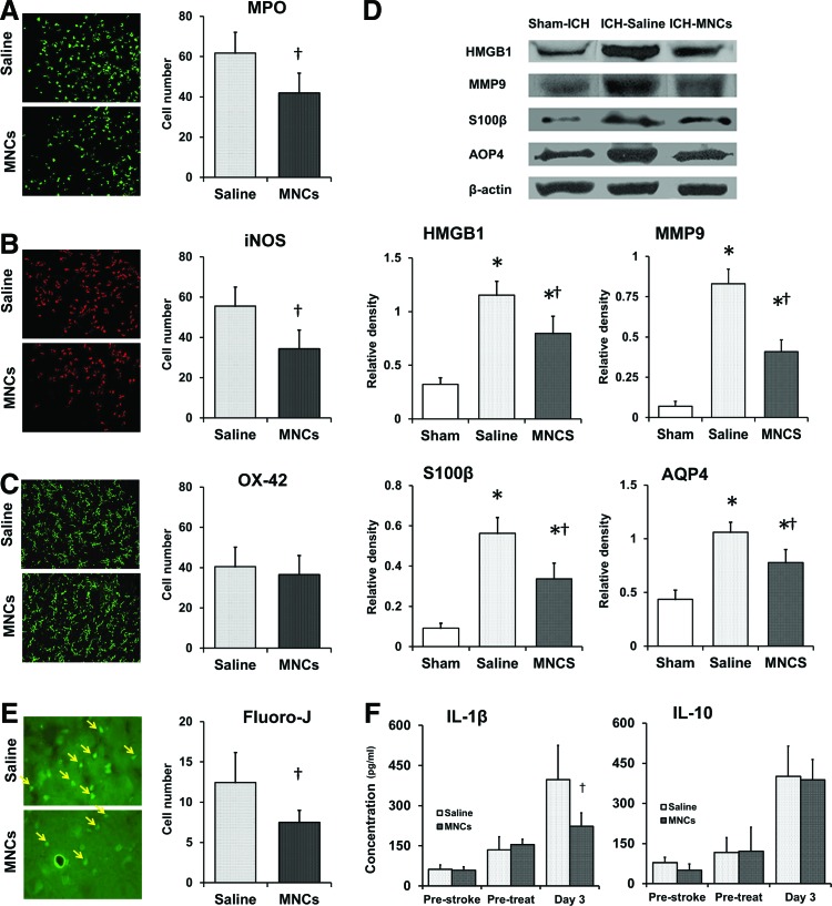
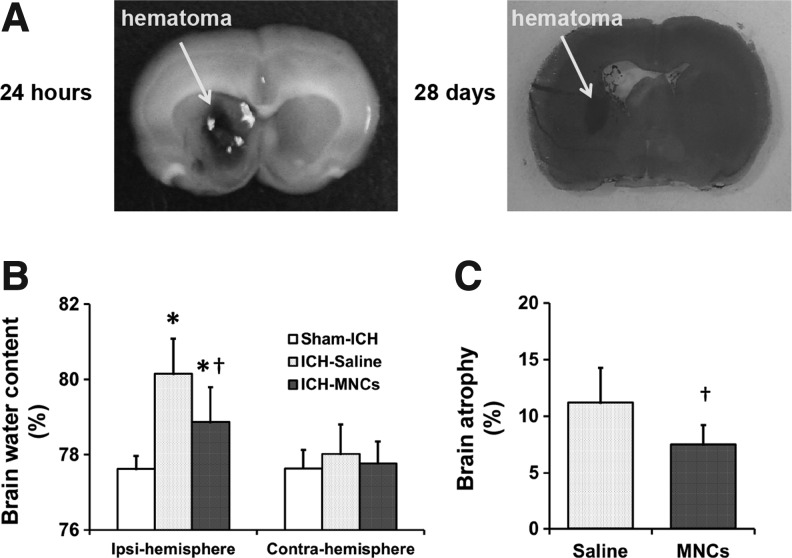
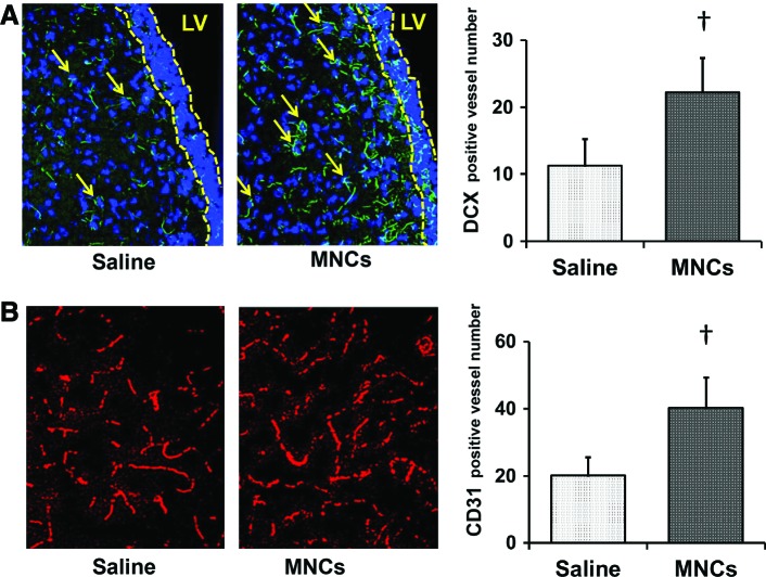
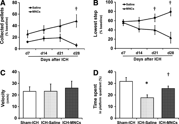
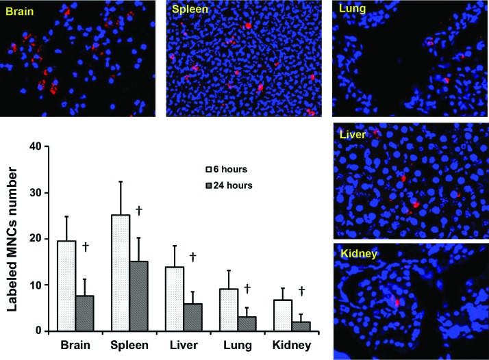
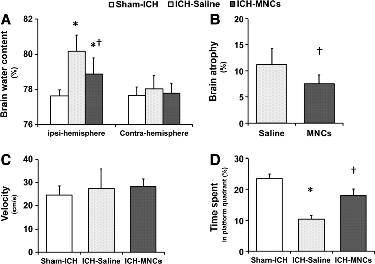
Autologous bone marrow-derived mononuclear cells (MNCs) are a potential therapy for ischemic stroke. However, the effect of MNCs in intracerebral hemorrhage (ICH) has not been fully studied. In this study, we investigated the effects of autologous MNCs in experimental ICH. ICH was induced by infusion of autologous blood into the left striatum in young and aged male Long Evans rats. Twenty-four hours after ICH, rats were randomized to receive an intravenous administration of autologous MNCs (1 × 10(7) cells/kg) or saline. We examined brain water content, various markers related to the integrity of the neurovascular unit and inflammation, neurological deficit, neuroregeneration, and brain atrophy. We found that MNC-treated young rats showed a reduction in the neurotrophil infiltration, the number of inducible nitric oxide synthase-positive cells, and the expression of inflammatory-related signalings such as the high-mobility group protein box-1, S100 calcium binding protein B, matrix metalloproteinase-9, and aquaporin 4. Ultimately, MNCs reduced brain edema in the perihematomal area compared with saline-treated animals at 3 days after ICH. Moreover, MNCs increased vessel density and migration of doublecortin-positive cells, improved motor functional recovery, spatial learning, and memory impairment, and reduced brain atrophy compared with saline-treated animals at 28 days after ICH. We also found that MNCs reduced brain edema and brain atrophy and improved spatial learning and memory in aged rats after ICH. We conclude that autologous MNCs can be safely harvested and intravenously reinfused in rodent ICH and may improve long-term structural and functional recovery after ICH. The results of this study may be applicable when considering future clinical trials testing MNCs for ICH.


Figures

<b>FIG. 1.</b>
FIG. 1.
Mononuclear cells (MNCs) modulate the inflammatory responses in the local brain after intracerebral hemorrhage (ICH). (A–C) MNCs lead to modulation of inflammatory cells that have infiltrated into the perihematomal area at day 3 after ICH, compared to saline-treated group: representative fluorescent photomicrographs show (A) myeloperoxidase (MPO)-positive cells (green), (B) inducible nitric oxide synthase (iNOS)-positive cells (red), and (C) OX-42-positive cells (green), and the right bar graphs illustrate the quantification analysis of MNC modulation, respectively. Data are the mean ± standard deviation (SD). P < 0.05, compared with saline-treated group after ICH. Magnification: 200×. n = 4 per group. (D) MNC treatment significantly ameliorated ICH-induced production of high-mobility group protein box-1 (HMGB1), S100 calcium binding protein B (S100β), matrix metalloproteinase-9 (MMP9), and aquaporin (AQP4) protein in the ICH-affected brain. The representative immunoblots and bar graphs show the relative quantitative expression levels of HMGB1, S100β, MMP9, and AQP4 protein in the hemorrhagic hemisphere at day 3 after ICH. Data are the mean ± SD. *P < 0.05 compared with sham-ICH-operated group. P < 0.05 compared with saline-treated group after ICH. n = 3 per group. (E) Representative fluorescent photomicrographs and bar graph show that MNC treatment significantly reduces the Fluoro-J-stained neuron number (yellow arrows) in the perihematomal area at day 3 after ICH, compared to saline control. Data are the mean ± SD. P < 0.05, compared with saline-treated group after ICH. Magnification: 630×. n = 4 per group. (F) Bar graphs indicate that MNC treatment modulates the cytokine level in the serum after ICH. Data are the mean ± SD. P < 0.05, compared with saline-treated group after ICH. n = 5 per group. Color images available online at www.liebertpub.com/scd
<b>FIG. 2.</b>
FIG. 2.
MNCs reduce perihematomal edema caused by ICH. (A) Representative pictures showed the hematomal position at 24 h and 28 days after autologous blood injection (from preliminary data). (B) Bar graphs illustrate the brain water content in the hemorrhagic (ipsilateral) and contralateral hemispheres at 3 days after ICH in the sham-, saline-, and MNC-treated ICH groups. Data are the mean ± SD. *P < 0.05 compared with the sham-ICH-operated group. P < 0.05 compared with saline-treated group after ICH. n = 6 per group. (C) Bar graphs illustrate that MNC treatment reduced brain atrophy at day 28 after ICH, compared with the saline-treated group. Data are the mean ± SD. P < 0.05 compared with the saline-treated group. n = 7 per group.
<b>FIG. 3.</b>
FIG. 3.
MNCs improved neurogenesis and angiogenesis at day 28 after ICH. (A) The representative photomicrographs showing the neuroblasts in the striatum near the subventricular zone. Only cells with the double doublecortin (DCX; green) and nuclear marker 4′,6-diamidino-2-phenylindole (DAPI) staining were enumerated as DCX-positive cells (arrows). The subventricular zone is shown as the area between the two broken yellow lines. Bar graph on the right shows the number of DCX-positive cells in the striatum in saline- and MNC-treated rats. P < 0.05, compared with the saline-treated group. Magnification: 200×. n = 4 per group. (B) Representative photomicrographs showing CD31-labeled blood vessels (red) at day 28 after stroke. Bar graphs on the right show the vessel numbers in saline- and MNC-treated rats. P < 0.05, compared with saline-treated group. Magnification: 200×. n = 4 per group. LV, lateral ventricle. Color images available online at www.liebertpub.com/scd
<b>FIG. 4.</b>
FIG. 4.
MNCs improved sensorimotor functional recovery after ICH. Sensorimotor function was evaluated by the staircase test: line diagrams indicate the alternation of the collected pellets on the right staircase (A) and the lowest step reached (B) in the staircase test over time from day 7 to 28 after ICH in rats after ICH treated with saline (control) or MNC. Data are the mean ± SD. P < 0.05, compared with the saline-treated group. n = 7 per group. Cognitive dysfunction at 4 weeks after ICH in sham group and in animals after ICH treated with saline or MNC was evaluated by the Morris water maze test using the percent time spent in the platform quadrant during the probe trial. The swimming speed was the same for all the groups (C). The bar graph shows time spent in the target quadrant by sham rats and rats after ICH treated with saline (control) or MNC. (D). Data are the mean ± SD. *P < 0.05 compared with sham-ICH-operated group. P = 0.071, compared with saline-treated group. Sham n = 13, saline n = 12, and MNCs n = 11.
<b>FIG. 5.</b>
FIG. 5.
MNC biodistributions at 6 and 24 h after IV administration in the ICH model. The representative fluorescence images and bar graph show the number of the Qtracker-labeled MNCs (red) in the brain, spleen, lungs, liver, and kidney. Red: Qtracker 655; blue: DAPI. Magnification: 200×. The y-axis is the mean number of cells collected from each section. Data are the mean ± SD. P < 0.05, compared with 6 h, n = 4 per time point. Color images available online at www.liebertpub.com/scd
<b>FIG. 6.</b>
FIG. 6.
The effect of MNCs on ICH outcome in retired bleeder Long Evans rat. (A) Bar graph illustrating the brain water content in the hemorrhagic (ipsilateral) and contralateral hemispheres. Data are the mean ± SD. *P < 0.05 compared with the sham-ICH-operated group. P < 0.05 compared with saline-treated group. n = 6 per group. (B) Bar graph illustrating that MNC reduced brain atrophy in animals at day 28 after ICH, compared with saline-treated controls. Data are the mean ± SD. P < 0.05 compared with saline-treated group. n = 7 per group. (C, D) The outcomes of Morris water maze test performed at 4 weeks after ICH. Bar graphs illustrate swimming speed (C) and the time spent in the target quadrant (D) in the sham-ICH group and ICH groups treated with saline or MNC. Data are the mean ± SD. *P < 0.05 compared with sham-ICH-operated group. P < 0.05 compared with saline-treated group. Sham n = 13, saline n = 12, and MNCs n = 10.

References

    1. Broderick J, Connolly S, Feldmann E, Hanley D, Kase C, et al. (2007). Guidelines for the management of spontaneous intracerebral hemorrhage in adults: 2007 update: a guideline from the American Heart Association/American Stroke Association Stroke Council, High Blood Pressure Research Council, and the Quality of Care and Outcomes in Research Interdisciplinary Working Group. Stroke 38:2001–2023 - PubMed
    1. Jorgensen HS, Nakayama H, Raaschou HO. and Olsen TS. (1995). Intracerebral hemorrhage versus infarction: stroke severity, risk factors, and prognosis. Ann Neurol 38:45–50 - PubMed
    1. Morgenstern LB, Hemphill JC, 3rd, Anderson C, Becker K, Broderick JP, et al. (2010). Guidelines for the management of spontaneous intracerebral hemorrhage: a guideline for healthcare professionals from the American Heart Association/American Stroke Association. Stroke 41:2108–2129 - PMC - PubMed
    1. Abe K, Yamashita T, Takizawa S, Kuroda S, Kinouchi H, et al. (2012). Stem cell therapy for cerebral ischemia: from basic science to clinical applications. J Cereb Blood Flow Metab 32:1317–1331 - PMC - PubMed
    1. Saito H, Magota K, Zhao S, Kubo N, Kuge Y, et al. (2013) 123I-iomazenil single photon emission computed tomography visualizes recovery of neuronal integrity by bone marrow stromal cell therapy in rat infarct brain. Stroke 44:2869–2874 - PubMed

Publication types

LinkOut - more resources