. 2016 Jan;30(1):122-7.
doi: 10.1089/end.2015.0298. Epub 2015 Oct 9.

A Proposed Grading System to Standardize the Description of Renal Papillary Appearance at the Time of Endoscopy in Patients with Nephrolithiasis

Affiliations

A Proposed Grading System to Standardize the Description of Renal Papillary Appearance at the Time of Endoscopy in Patients with Nephrolithiasis

Michael S Borofsky et al. J Endourol. 2016 Jan.

Abstract

Background and purpose: The appearance of the renal papillae in patients with nephrolithiasis can be quite variable and can range from entirely healthy to markedly diseased. The implications of such findings remain unknown. One potential reason is the lack of a standardized system to describe such features. We propose a novel grading scale to describe papillary appearance at the time of renal endoscopy.

Methods: Comprehensive endoscopic renal assessment and mapping were performed on more than 300 patients with nephrolithiasis. Recurring abnormal papillary characteristics were identified and quantified based on degree of severity.

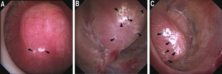
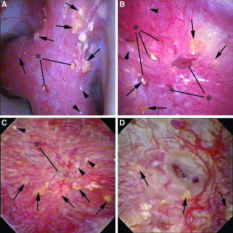
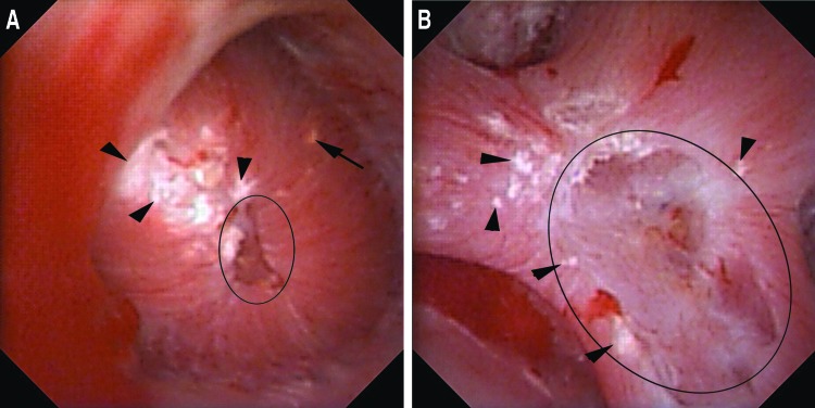
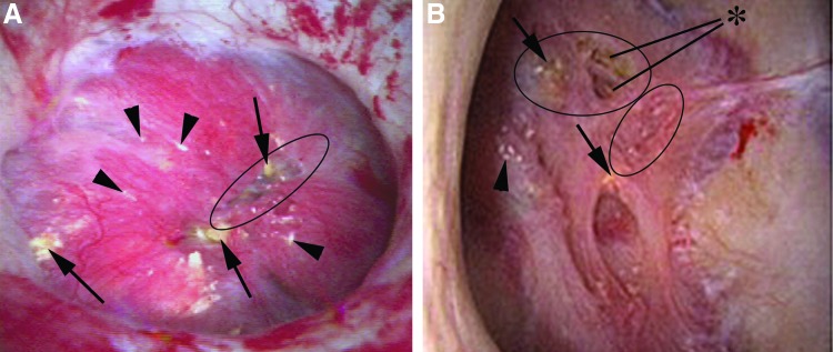
Results: Four unique papillary features were chosen for inclusion in the PPLA scoring system- ductal Plugging, Pitting, Loss of contour, and Amount of Randall's plaque. Unique scores are calculated for individual papillae based on reference examples.

Conclusions: The description and study of renal papillary appearance in stone formers have considerable potential as both a clinical and research tool; however, a standardized grading system is necessary before using it for these purposes.


Figures

<b>FIG. 1.</b>
FIG. 1.
(A) Healthy papilla with mild amount of Randall's plaque (black arrowheads) (Plugging = 0, Pitting = 0, Loss of Contour = 0, Randall's plaque = a [PPLA 0a]). (B) Healthy papilla with moderate amount of Randall's plaque (black arrowheads) (Plugging = 0, Pitting = 0, Loss of Contour = 0, Randall's plaque = b [PPLA 0b]). (C) Healthy papilla with extensive amount of Randall's plaque (black arrowheads) (Plugging = 0, Pitting = 0, Loss of Contour = 0, Randall's plaque = c [PPLA 0c]).
<b>FIG. 2.</b>
FIG. 2.
(A) This compound papilla demonstrates numerous deposits of yellow plaque, cumulatively more than 5 (black arrows). Dilated ducts with protruding yellow mineral deposits are designated with the asterisk. There is no evidence of pitting or loss of contour. A minute amount of Randall's plaque can be visualized by the arrowheads in the lower right corner (Plugging = 2, Pitting = 0, Loss of Contour = 0, Randall's plaque = a [PPLA 2a]). (B) This papilla also demonstrates plugging as evidenced by both yellow plaque (black arrows) and dilated ducts, some of which contain protruding yellow mineral deposits (asterisk). There is no evidence of pitting and an intermediate loss of contour. A minimal amount of Randall's plaque can be seen as designated by the black arrowheads (Plugging = 2, Pitting = 0, Loss of Contour = 1, Randall's plaque = a (PPLA 3a). (C) This papilla has many yellow plaque deposits (black arrows). One large dilated duct can be seen in the center of the papilla (asterisk) that is magnified in Figure 2D. There is no pitting, intermediate loss of contour, and minimal Randall's plaque (Plugging = 2, Pitting = 0, Loss of Contour = 1, Randall's plaque = a [PPLA 3a]). (D) This image is the magnification of the central region of Figure 2C. Yellow plaque deposits are again designated with black arrows. The prominent dilated duct of Bellini is designated by the asterisk within it.
<b>FIG. 3.</b>
FIG. 3.
(A) This papilla has pitting focused at the periphery of the papillary tip (black circle). Less than 25% of the papillary surface is affected. A single yellow plaque deposit is marked by a black arrow. The papilla has no loss of contour and a severe amount of Randall's plaque (black arrowheads) (Plugging = 1, Pitting = 1, Loss of Contour = 0, Randall's plaque = c [PPLA 2c]). (B) This compound papilla demonstrates pitting with more than 25% of the papillary surface affected (black circle). There is no evidence of plugging or loss of contour. Moderate Randall's plaque is seen (Plugging = 0, Pitting = 2, Loss of Contour = 0, Randall's plaque = b [PPLA 2b]).
<b>FIG. 4.</b>
FIG. 4.
(A) This papilla has a number of abnormal findings including numerous deposits of yellow plaque (black arrows), <25% pitting (black circle), a moderate loss of contour, and several small foci of Randall's plaque (black arrowheads) (Plugging = 2, Pitting = 1, Loss of Contour = 1, Randall's plaque = a [PPLA 4a]). (B) This papilla has several foci of yellow plaque (black arrows) and dilated ducts (asterisk), moderate pitting (black circle), and complete loss of contour. There is minimal Randall's plaque (arrowhead) (Plugging = 2, Pitting = 1, Loss of Contour = 2, Randall's plaque = a [PPLA 5a]).

References

    1. Evan AP, Lingeman J, Coe F, et al. . Renal histopathology of stone-forming patients with distal renal tubular acidosis. Kidney Int 2007;71:795–801 - PubMed
    1. Matlaga BR, Williams JC, Jr, Kim SC, et al. . Endoscopic evidence of calculus attachment to Randall's plaque. J Urol 2006;175:1720–1724 - PubMed
    1. Miller NL, Williams JC, Jr, Evan AP, et al. . In idiopathic calcium oxalate stone-formers, unattached stones show evidence of having originated as attached stones on Randalls plaque. BJU Int 2010;105:242–245 - PMC - PubMed
    1. Coe FL, Evan AP, Worcester EM, Lingeman JE. Three pathways for human kidney stone formation. Urol Res 2010;38:147–160 - PMC - PubMed
    1. Evan AE, Lingeman JE, Coe FL, et al. . Histopathology and surgical anatomy of patients with primary hyperparathyroidism and calcium phosphate stones. Kidney Int 2008;74:223–229 - PubMed

Publication types

LinkOut - more resources