. 2016 Apr 1;33(7):625-40.
doi: 10.1089/neu.2015.3970. Epub 2015 Nov 23.

Altered Neuroinflammation and Behavior after Traumatic Brain Injury in a Mouse Model of Alzheimer's Disease

Affiliations

Altered Neuroinflammation and Behavior after Traumatic Brain Injury in a Mouse Model of Alzheimer's Disease

Olga Kokiko-Cochran et al. J Neurotrauma. .

Abstract

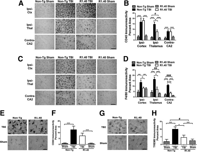
Traumatic brain injury (TBI) has acute and chronic sequelae, including an increased risk for the development of Alzheimer's disease (AD). TBI-associated neuroinflammation is characterized by activation of brain-resident microglia and infiltration of monocytes; however, recent studies have implicated beta-amyloid as a major manipulator of the inflammatory response. To examine neuroinflammation after TBI and development of AD-like features, these studies examined the effects of TBI in the presence and absence of beta-amyloid. The R1.40 mouse model of cerebral amyloidosis was used, with a focus on time points well before robust AD pathologies. Unexpectedly, in R1.40 mice, the acute neuroinflammatory response to TBI was strikingly muted, with reduced numbers of CNS myeloid cells acquiring a macrophage phenotype and decreased expression of inflammatory cytokines. At chronic time points, macrophage activation substantially declined in non-Tg TBI mice; however, it was relatively unchanged in R1.40 TBI mice. The persistent inflammatory response coincided with significant tissue loss between 3 and 120 days post-injury in R1.40 TBI mice, which was not observed in non-Tg TBI mice. Surprisingly, inflammatory cytokine expression was enhanced in R1.40 mice compared with non-Tg mice, regardless of injury group. Although R1.40 TBI mice demonstrated task-specific deficits in cognition, overall functional recovery was similar to non-Tg TBI mice. These findings suggest that accumulating beta-amyloid leads to an altered post-injury macrophage response at acute and chronic time points. Together, these studies emphasize the role of post-injury neuroinflammation in regulating long-term sequelae after TBI and also support recent studies implicating beta-amyloid as an immunomodulator.

Keywords: Alzheimer's disease; macrophage; neuroinflammation; traumatic brain injury.


Figures

<b>FIG. 1.</b>
FIG. 1.
Lesion and hippocampal volume change over time. (A) Representative three-dimensional (3D) reconstructions show a cortical injury cavity at 3 days post-injury (DPI) in mice with traumatic brain injury (TBI), which is larger at 120 DPI (n = 6-7/group/time point). (B) Volumetric lesion analysis reveals a time-dependent increase in the size of the injury cavity in the R1.40 mice at 120 DPI compared with 3 DPI. (C) Representative 3D reconstructions show the ipsilateral hippocampus in brain and sham injured non-Tg and R1.40 mice at 3 and 120 DPI. (D) Volumetric hippocampal analysis reveals a time-dependent decrease in all groups, which reached statistical significance in R1.40 TBI and sham mice.
<b>FIG. 2.</b>
FIG. 2.
Traumatic brain injury (TBI) induces morphometric changes in Iba1+ macropahges. (A,C) Representative images of Iba1 immunostaining in sham and TBI mice in ipsilateral cortex (Ipsi-Ctx), ipsilateral thalamus (Ipsi-Thal), and contralateral CA2 (Contra-CA2) brain regions at 3 days post-injury (DPI) and 120 DPI, respectively. (B) Form factor (FF) analysis of Iba1 positive macrophages at 3 DPI revealed higher values in non-Tg and R1.40 TBI mice compared with sham mice in all brain regions analyzed (n = 6/group). (C) FF analysis at 120 DPI also revealed a primary effect of injury in all brain regions (n = 6/ group). Error bars indicate standard error of the mean. Scale bars indicate 20 μm. Main effect of injury, *, p < 0.05, ***, p < 0.001; main effect of genotype, #, p < 0.05.
<b>FIG. 3.</b>
FIG. 3.
Distinct macrophage phenotype in brain injured R1.40 mice at 3 days post-injury (DPI). (A,C) Representative images of CD45 and F4/80 immunostaining in sham and mice with traumatic brain injury (TBI) in ipsilateral cortex (Ipsi-Ctx), ipsilateral thalamus (Ipsi-Thal), and contralateral CA2 (Contra-CA2) brain regions at 3 DPI. (B) Non-Tg and R1.40 TBI mice show similar levels of increased CD45 immunoreactivity compared with sham animals (n = 6/group) in all brain regions. R1.40 TBI mice showed a decrease in CD45 immunoreactivity compared with non-Tg TBI mice in the Ipsi-Thal. (D) F4/80 immunoreactivity was significantly elevated in non-Tg TBI mice compared with all other groups in each brain region analyzed (n = 6/group). (E,G) Representative images of CD68 and Trem2 immunostaining in sham and TBI mice at 3 DPI. (F) CD68 expression was significantly increased in TBI mice compared with shams with non-Tg TBI mice exhibiting the most CD68 expression (n = 3/group). (H) Trem2 expression was significantly elevated in non-Tg TBI mice compared with all other groups (n = 3/group). Error bars indicate standard error of the mean. Scale bars indicate 20 μm. Main effect of injury: **, p < 0.01; ***, p < 0.001; Main effect of genotype: #, p < 0.05; ##, p < 0.01; ###, p < 0.001; interaction effect: +, p < 0.05; +++, p < 0.001.
<b>FIG. 4.</b>
FIG. 4.
Proportion of microglia and monocytes are similar between non-Tg and R1.40 mice with traumatic brain injury (TBI). (A,B) Representative contour plots of microglia (CD45low/F4/80 cells) and monocytes (CD45high/F4/80 cells) in non-Tg and R1.40 mice with TBI at 3 days post-injury (DPI). (C) R1.40 TBI mice show a significant decrease in CD45+/F4/80+ compared with non-Tg TBI mice at 3 DPI (n = 3–7/group). (D,E) TBI induced a significant reduction in microglia (CD45low/F4/80 cells) at 3 DPI; however, no significant differences in monocytes were identified at 3 DPI (CD45high/F4/80 cells). Error bars indicate standard error of the mean; main effect of injury: *, p < 0.05; interaction effect: +, p < 0.05.
<b>FIG. 5.</b>
FIG. 5.
Macrophage activation persists in subcortical structures at 120 days post-injury (DPI). (A,C) Representative images of CD45 and F4/80 immunostaining of both non-Tg and R1.40 sham and mice with traumatic brain injury (TBI) at 120 DPI. (B) CD45 immunoreactivity remained elevated in the ipsilateral thalamus (Ipsi-Thal) in both non-Tg and R1.40 TBI mice (n = 6/group). (D) A main effect of injury was apparent in F4/80 immunoreactivity in the Ipsi-Thal and contralateral CA2 (Contra-CA2) region. Error bars indicate standard error of the mean. Scale bars indicate 20 μm. Main effect of injury: *, p < 0.05, **; p < 0.01. Ipsilateral cortex, Ipsi-Ctx.
<b>FIG. 6.</b>
FIG. 6.
Traumatic brain injury (TBI) causes time dependent changes in APP expression. (A,D) Western blot of cortical extracts were probed with antibodies CT15, 6E10, and GAPDH at 3 and 120 days post-injury (DPI) respectively. (B) There was a significant increase in APP in non-Tg TBI mice compared with non-Tg shams at 3 DPI. R1.40 TBI mice displayed higher levels of APP than non-Tg TBI mice, but no differences in APP expression were identified between R1.40 TBI and R1.40 sham mice at 3 DPI. (C) No difference in human APP expression was found between R1.40 TBI and R1.40 sham mice at 3 DPI. As expected, human APP was undetectable in non-Tg mice. (E) There was a significant reduction in APP in non-Tg TBI mice compared with non-Tg shams at 120 DPI. APP levels were reduced in non-Tg mice compared with R1.40 mice regardless of injury group. (F) R1.40 TBI mice displayed a significant reduction in human APP expression compared with R1.40 sham mice at 120 DPI (3 DPI, n = 5–6/group; 120 DPI, n = 6/group). Human APP was undetectable in non-Tg mice. (G,H) Levels of Aβ 1-40 in ipsilateral cortex homogenates were higher in R1.40 mice compared with non-Tg mice at 3 and 120 DPI, respectively; however these differences were not statistically significant (n = 5–6/group). (I) Representative images of CT15 immunostaining in sham and TBI mice at 3 DPI (top two rows) and 120 DPI (bottom row). APP accumulation was apparent in the ipsilateral external capsule and cortex at 3 DPI in mice with TBI. By 120 DPI, only R1.40 mice showed enhanced APP immunostaining in the ipsi-cortex (n = 3/group/time point). Error bars indicate standard error of the mean. Scale bars indicate 20 μm. Main effect of injury: *, p < 0.05; main effect of genotype: #, p < 0.05; ###, p < 0.001.
<b>FIG. 7.</b>
FIG. 7.
Traumatic brain injury (TBI) induces time dependent changes in motor and cognitive function. (A) Mean latency to fall from the rotating rod was shorter in TBI mice at all post-injury time points compared to shams regardless of genotype. No between genotype differences in rotarod performance were detected in sham and TBI mice (n = 12–14/group). (B) No significant differences between groups in average spontaneous alternation in the y maze. R1.40 TBI mice showed a significant reduction in percent spontaneous alternation between arms from 7 days post-injury (DPI) to 90 DPI. (C) Average total arm entries were similar between TBI groups; however, sham mice demonstrated a significant reduction in arm entries over time (n = 12–14/group). (D) A significant interaction effect between injury and genotype was detected in latency to reach the hidden platform at 120 DPI (n = 12–14/group). (E) Average acquisition index shows that latency to reach the platform significantly decreases within a testing day in R1.40 compared with non-Tg mice. (F) Average savings index shows that latency to reach the platform significantly increases between testing days in R1.40 compared with Non-Tg mice. Error bars indicate standard error of the mean. Main effect of injury: *, p < 0.05; ***, p < 0.001; Interaction effect: ++, p < 0.01; +++, p < 0.001.

References

    1. Nemetz P.N., Leibson C., Naessens J.M., Beard M., Kokmen E., Annegers J.F., and Kurland L.T. (1999). Traumatic brain injury and time to onset of Alzheimer's disease: a population-based study. Am. J. Epidemiol. 149, 32–40 - PubMed
    1. Schofield P.W., Tang M., Marder K., Bell K., Dooneief G., Chun M., Sano M., Stern Y., and Mayeux R. (1997). Alzheimer's disease after remote head injury: an incidence study. J. Neurol. Neurosurg. Psychiatry 62, 119–124 - PMC - PubMed
    1. Fleminger S., Oliver D.L., Lovestone S., Rabe-Hesketh S., and Giora A. (2003). Head injury as a risk factor for Alzheimer's disease: the evidence 10 years on; a partial replication. J. Neurol. Neurosurg. Psychiatry 74, 857–862 - PMC - PubMed
    1. Plassman B.L., Havlik R.J., Steffens D.C., Helms M.J., Newman T.N., Drosdick D., Phillips C., Gau B.A., Welsh-Bohmer K.A., Burke J.R., Guralnik J.M., and Breitner J.C. (2000). Documented head injury in early adulthood and risk of Alzheimer's disease and other dementias. Neurology 55, 1158–1166 - PubMed
    1. Mortimer J.A., van Duijn C.M., Chandra V., Fratiglioni L., Graves A.B., Heyman A., Jorm A.F., Kokmen E., Kondo K., Rocca W.A., Shalat S.L., Soininen H., and Hofman A. (1991). Head trauma as a risk factor for Alzheimer's disease: a collaborative re-analysis of case-control studies. EURODEM Risk Factors Research Group. Int. J. Epidemiol. 20, Suppl 2, S28–S35 - PubMed

Publication types

MeSH terms

Substances

LinkOut - more resources