

F429 Regulation of Tunnels in Cytochrome P450 2B4: A Top Down Study of Multiple Molecular Dynamics Simulations
- PMID: 26415031
- PMCID: PMC4587367
- DOI: 10.1371/journal.pone.0137075
F429 Regulation of Tunnels in Cytochrome P450 2B4: A Top Down Study of Multiple Molecular Dynamics Simulations
Abstract
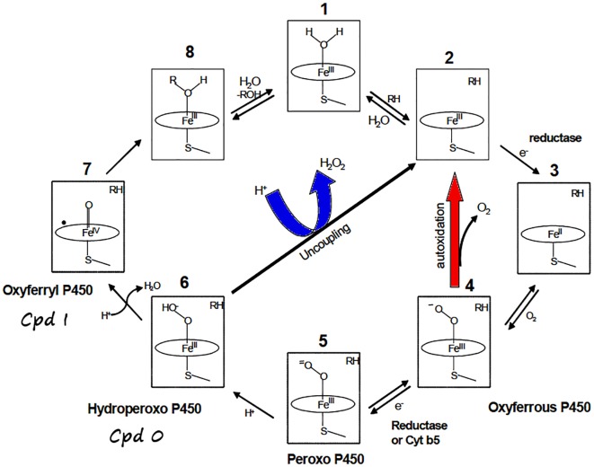
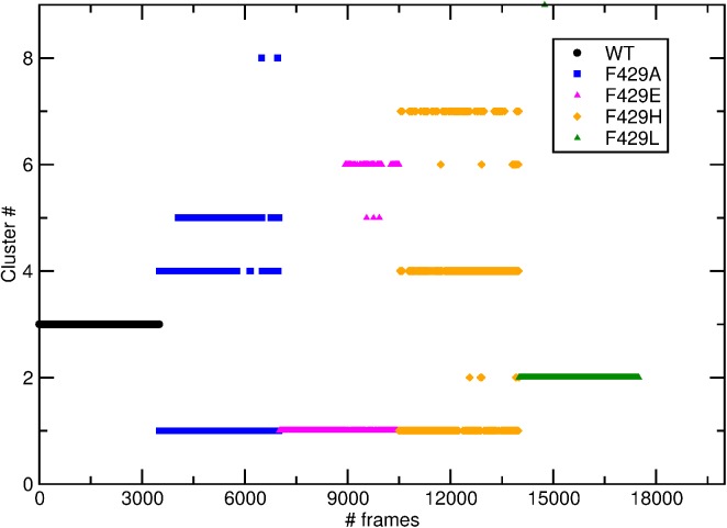
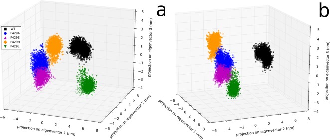
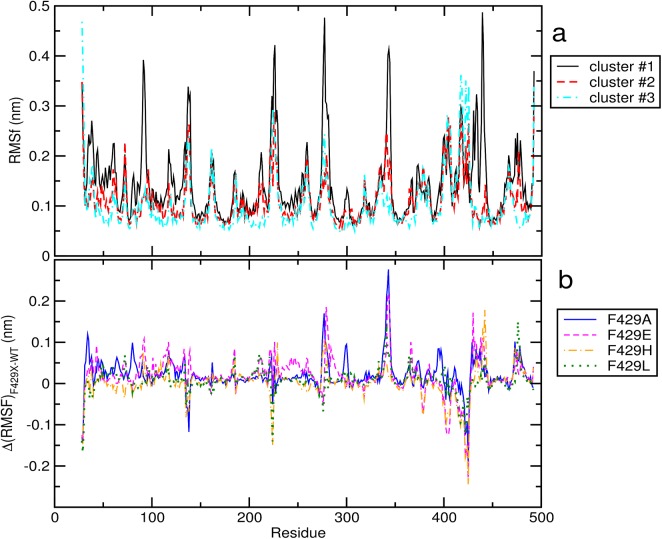
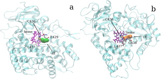
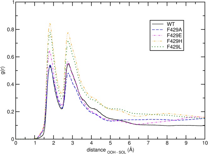
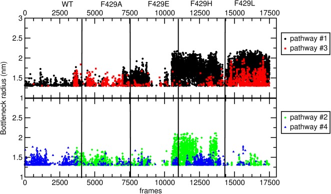
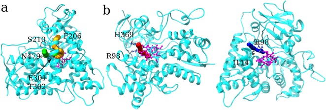
The root causes of the outcomes of the single-site mutation in enzymes remain by and large not well understood. This is the case of the F429H mutant of the cytochrome P450 (CYP) 2B4 enzyme where the substitution, on the proximal surface of the active site, of a conserved phenylalanine 429 residue with histidine seems to hamper the formation of the active species, Compound I (porphyrin cation radical-Fe(IV) = O, Cpd I) from the ferric hydroperoxo (Fe(III)OOH-, Cpd 0) precursor. Here we report a study based on extensive molecular dynamic (MD) simulations of 4 CYP-2B4 point mutations compared to the WT enzyme, having the goal of better clarifying the importance of the proximal Phe429 residue on CYP 2B4 catalytic properties. To consolidate the huge amount of data coming from five simulations and extract the most distinct structural features of the five species studied we made an extensive use of cluster analysis. The results show that all studied single polymorphisms of F429, with different side chain properties: i) drastically alter the reservoir of conformations accessible by the protein, perturbing global dynamics ii) expose the thiolate group of residue Cys436 to the solvent, altering the electronic properties of Cpd0 and iii) affect the various ingress and egress channels connecting the distal sites with the bulk environment, altering the reversibility of these channels. In particular, it was observed that the wild type enzyme exhibits unique structural features as compared to all mutant species in terms of weak interactions (hydrogen bonds) that generate a completely different dynamical behavior of the complete system. Albeit not conclusive, the current computational investigation sheds some light on the subtle and critical effects that proximal single-site mutations can exert on the functional mechanisms of human microsomal CYPs which should go rather far beyond local structure characterization.
Conflict of interest statement
Figures


Similar articles
-
Role of the Proximal Cysteine Hydrogen Bonding Interaction in Cytochrome P450 2B4 Studied by Cryoreduction, Electron Paramagnetic Resonance, and Electron-Nuclear Double Resonance Spectroscopy.Biochemistry. 2016 Feb 16;55(6):869-83. doi: 10.1021/acs.biochem.5b00744. Epub 2016 Feb 3. Biochemistry. 2016. PMID: 26750753 Free PMC article.
-
Structural and functional characterization of a cytochrome P450 2B4 F429H mutant with an axial thiolate-histidine hydrogen bond.Biochemistry. 2014 Aug 12;53(31):5080-91. doi: 10.1021/bi5003794. Epub 2014 Jul 31. Biochemistry. 2014. PMID: 25029089 Free PMC article.
-
Multiresolution molecular dynamics simulations reveal the interplay between conformational variability and functional interactions in membrane-bound cytochrome P450 2B4.Protein Sci. 2024 Oct;33(10):e5165. doi: 10.1002/pro.5165. Protein Sci. 2024. PMID: 39291728 Free PMC article.
-
Role of cytochrome b5 in catalysis by cytochrome P450 2B4.Biochem Biophys Res Commun. 2005 Dec 9;338(1):499-506. doi: 10.1016/j.bbrc.2005.09.022. Epub 2005 Sep 15. Biochem Biophys Res Commun. 2005. PMID: 16182240 Review.
-
Targeting of the highly conserved threonine 302 residue of cytochromes P450 2B family during mechanism-based inactivation by aryl acetylenes.Arch Biochem Biophys. 2011 Mar 1;507(1):135-43. doi: 10.1016/j.abb.2010.09.006. Epub 2010 Sep 15. Arch Biochem Biophys. 2011. PMID: 20836985 Free PMC article. Review.
Cited by
-
Molecular Dynamics Simulations Enforcing Nonperiodic Boundary Conditions: New Developments and Application to the Solvent Shifts of Nitroxide Magnetic Parameters.J Chem Theory Comput. 2022 Apr 12;18(4):2479-2493. doi: 10.1021/acs.jctc.2c00046. Epub 2022 Mar 8. J Chem Theory Comput. 2022. PMID: 35257572 Free PMC article.
-
Immersive virtual reality in computational chemistry: Applications to the analysis of QM and MM data.Int J Quantum Chem. 2016 Nov 15;116(22):1731-1746. doi: 10.1002/qua.25207. Epub 2016 Jul 13. Int J Quantum Chem. 2016. PMID: 27867214 Free PMC article.
-
Variability of the Cyclin-Dependent Kinase 2 Flexibility Without Significant Change in the Initial Conformation of the Protein or Its Environment; a Computational Study.Iran J Biotechnol. 2016 Jun;14(2):1-12. doi: 10.15171/ijb.1419. Iran J Biotechnol. 2016. PMID: 28959320 Free PMC article.
-
Computational Spectroscopy in Solution by Integration of Variational and Perturbative Approaches on Top of Clusterized Molecular Dynamics.J Chem Theory Comput. 2020 Sep 8;16(9):5747-5761. doi: 10.1021/acs.jctc.0c00454. Epub 2020 Aug 11. J Chem Theory Comput. 2020. PMID: 32697580 Free PMC article.
-
Structure-function relationship between soluble epoxide hydrolases structure and their tunnel network.Comput Struct Biotechnol J. 2021 Dec 13;20:193-205. doi: 10.1016/j.csbj.2021.10.042. eCollection 2022. Comput Struct Biotechnol J. 2021. PMID: 35024092 Free PMC article.
References
-
- Ortiz de Montellano PR, editor. Cytochrome P450: structure, mechanism, and biochemistry 3rd ed. New York: Kluwer Academic/Plenum Publishers; 2005.
Publication types
MeSH terms
Substances
LinkOut - more resources
Full Text Sources
Other Literature Sources
