. 2015 Sep 29:16:118.
doi: 10.1186/s12931-015-0278-3.

BARD1 mediates TGF-β signaling in pulmonary fibrosis

Affiliations

BARD1 mediates TGF-β signaling in pulmonary fibrosis

Pierre-Alain André et al. Respir Res. .

Abstract

Background: Idiopathic pulmonary fibrosis (IPF) is a rapid progressive fibro-proliferative disorder with poor prognosis similar to lung cancer. The pathogenesis of IPF is uncertain, but loss of epithelial cells and fibroblast proliferation are thought to be central processes. Previous reports have shown that BARD1 expression is upregulated in response to hypoxia and associated with TGF-β signaling, both recognized factors driving lung fibrosis. Differentially spliced BARD1 isoforms, in particular BARD1β, are oncogenic drivers of proliferation in cancers of various origins. We therefore hypothesized that BARD1 and/or its isoforms might play a role in lung fibrosis.

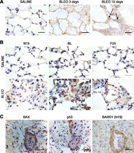
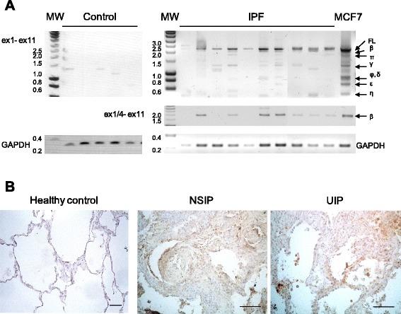
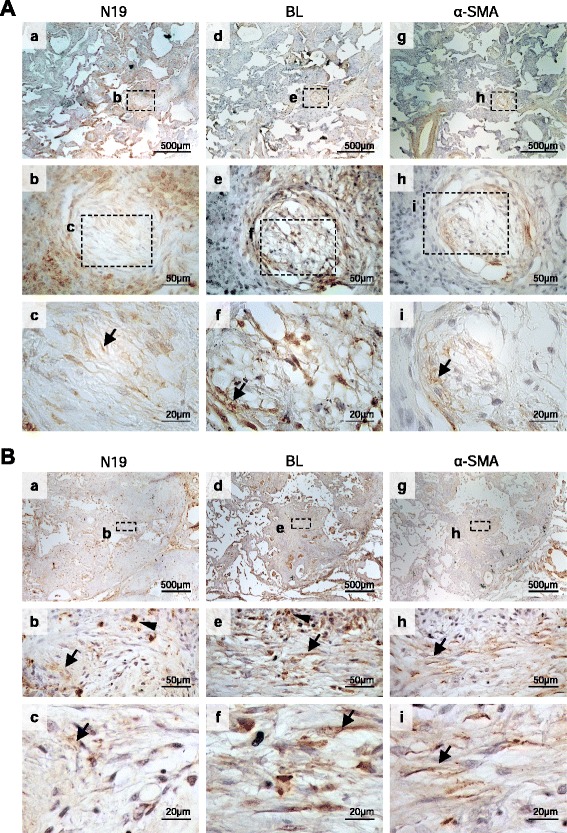
Methods: We investigated BARD1 expression as a function of TGF-β in cultured cells, in mice with experimentally induced lung fibrosis, and in lung biopsies from pulmonary fibrosis patients.

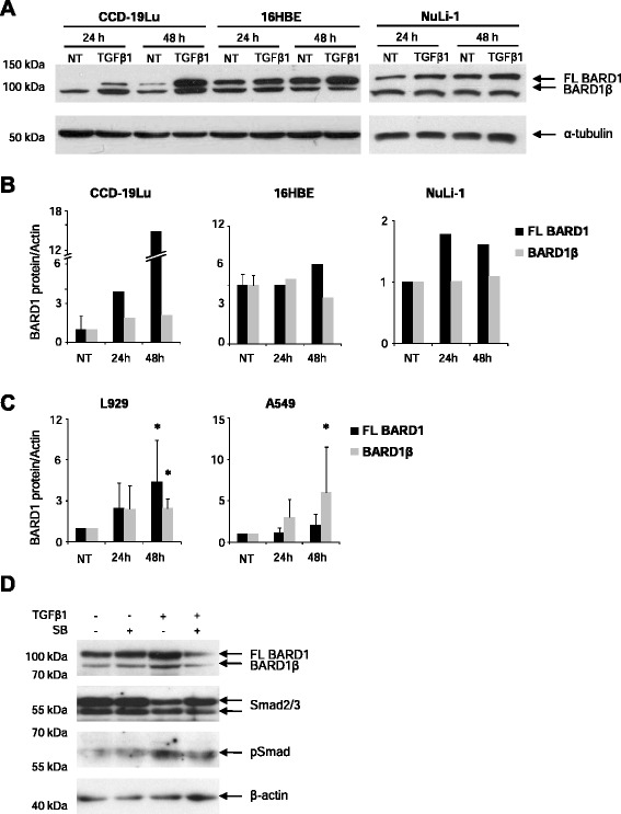
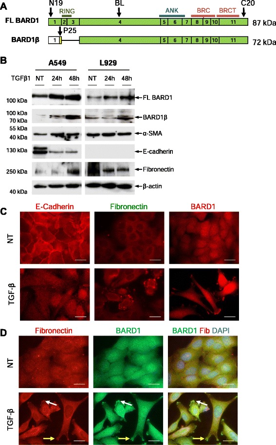
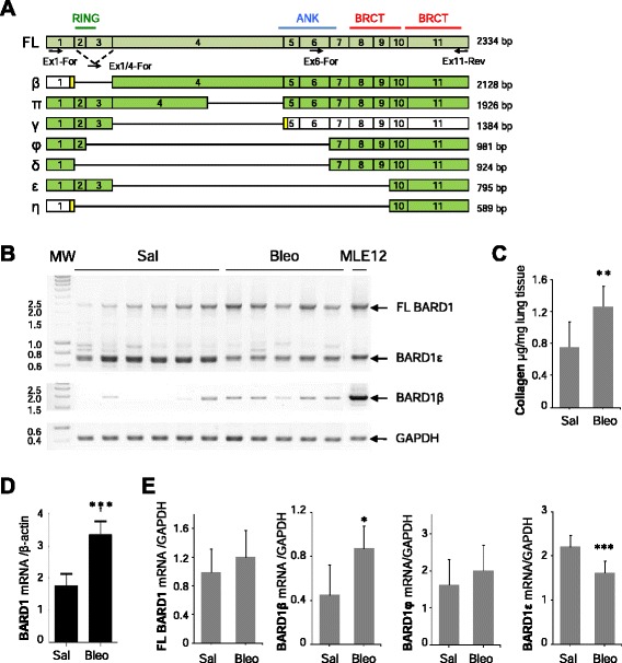
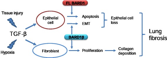
Results: FL BARD1 and BARD1β were upregulated in response to TGF-β in epithelial cells and fibroblasts in vitro and in vivo. Protein and mRNA expression studies showed very low expression in healthy lung tissues, but upregulated expression of full length (FL) BARD1 and BARD1β in fibrotic tissues.

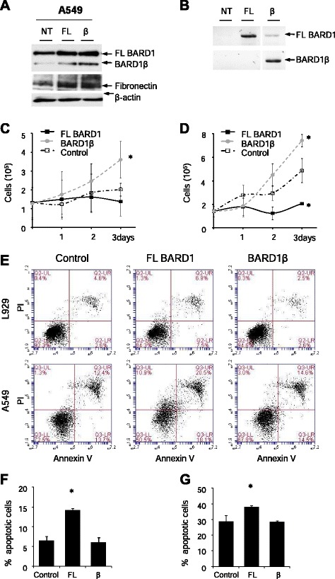
Conclusion: Our data suggest that FL BARD1 and BARD1β might be mediators of pleiotropic effects of TGF-β. In particular BARD1β might be a driver of proliferation and of pulmonary fibrosis pathogenesis and progression and represent a target for treatment.


Figures

Fig. 1
Fig. 1
TGF-β modulates BARD1 expression. Human lung fibroblasts (CCD-19Lu), bronchial epithelial cells (16HBE, NuLi-1), alveolar basal epithelial cells (A549), and mouse fibroblasts (L929) were cultured in absence (NT) or presence of TGF-β1 (10 ng/ml), and cell extracts were prepared after 24, and 48 h of treatment. a Western Blots of CCD-19Lu, 16HBE and NuLi-1 cell extracts prepared at 24 and 48 h of TGF-β treatment, probed with anti-BARD1 BL (mapping exon 4 of BARD1) antibody recognizing FL BARD1 and BARD1β. b CCD19-Lu and 16HBE and (c) A549 and L929 Western Blots were quantified by measuring spot density using AlphaEaseFc software. Each bar represents mean + SD (n ≥2 experiments); two-tailed Student’s T-Test *p < 0.05, TGF-β1 versus non-treated. d A549 culture in presence of SB-431542 1 h prior TGF-β1 treatment. Cell extracts were prepared after 48 h of TGF-β1/SB-431542 treatment and probed with anti-BARD1 BL, anti-Smad2/3, anti-pSmad antibodies
Fig. 2
Fig. 2
TGF-β induces BARD1β overexpression and localization. a Schematic representation of cDNA structure of FL BARD1 and N-terminally truncated isoform BARD1β. Approximate locations of protein motifs RING finger (RING), ankyrin repeats (ANK), and BRCT domains (BRCT) of BARD1 are indicated. Green exons indicate protein coding open reading frame (ORF). BARD1β encodes an alternative ORF in the first exon. Arrows indicated approximate positions of epitopes reactive with the antibodies used. b Lung epithelial cells (A549) and fibroblasts (L929) were cultured in absence (NT) or presence of TGF-β1, and cell extracts were prepared after 24 and 48 h. Western blots of cell extracts were probed with anti-BARD1 BL (exon 4), BARD1β-specific P25 (alternative ORF of BARD1β), α-SMA, E-cadherin, fibronectin, and β-actin antibodies. c A549 were treated with TGF-β1 and cells were fixed after 48 h. Immunofluoresence was performed with anti-E-cadherin, fibronectin, or BARD1 BL (exon 4) antibodies. d Co-immunostaining with anti-BARD1 BL and fibronectin (Fib) antibodies. TGF-β1 modulates BARD1 localization to areas at the cell membrane (yellow arrows) and cytoplasmic vesicles (white arrows), similar to and co-staining with fibronectin. Scale bars 20 μm
Fig. 3
Fig. 3
Differential effect of FL BARD1 and BARD1β on cell proliferation and apoptosis. a-b A549 cells were transfected with FL BARD1 and BARD1β expressing plasmids and cell extracts were prepared after 48 h. a Western Blots probed with anti-BARD1 (BL) and fibronectin. b RT-PCR analysis performed with primers for amplification of full length (FL) BARD1 (exon 1 to exon 11) or isoform BARD1β using primers from exon 1/4 junction and exon 11. c A549 and (d) L929 cell proliferation of control transformed cells (pcDNA), and cells expressing exogenous FL BARD1 or BARD1β was monitored over 3 days. BARD1β leads to increased proliferation, as described before. e Annexin V and PI staining of (e, f) A549 and (e, g) L929 24 h after transfection with FL BARD1, BARD1β or pcDNA control expressing plasmids. FL BARD1 induced cell apoptosis. Each bar represents mean + SD (n ≥2 experiments); two-tailed Student’s T-Test *p < 0.05, BARD1 versus control
Fig. 4
Fig. 4
RNA expression pattern of BARD1 mRNA isoforms in bleomycin induced lung fibrosis. a Exon structures of mRNAs of FL BARD1 and isoforms are aligned. Locations of protein motifs are indicated as in Fig. 2a. Greek names of isoforms are indicated on the left and size in bp on the right. Exons with open reading frames (ORF) are marked as green, non-coding as white, alternative ORFs as yellow. Arrows indicate position of forward (For) and reversed (Rev) primers used for RT-PCR. b RT-PCR on lung tissues from control (Saline) and Bleomycin (Bleo)-treated mice at 15 days after treatment is shown, performed with primers amplifying exon 1 to 11 or the region from exon 1/4 junction (BARD1β-specific) to exon 11 (ex1/4- ex11). Amplicons of BARD1 isoforms are indicated with Greek letters. GAPDH was amplified as RNA quantity and quality control. c Collagen expression was determined by Sircol assay at 15 days after bleomycin treatment. d Quantitative PCR analysis at 15 days after bleomycin assay showed a significant increase of BARD1 expression. e Semi-quantitative RT-PCR analysis at 15 days after bleomycin treatment of FL BARD1, BARD1β, BARD1φ, and BARD1ε mRNA expression showed a significant increase of BARD1β expression and a highly significant decrease of BARD1ε. Each bar represents mean + SD (n ≥6 mice in each group); Student’s T-Test *p < 0.05, **p < 0.01, ***p < 0.005 Bleo- versus saline-treated
Fig. 5
Fig. 5
BARD1 epitopes differential expression and its association with apoptosis in in bleomycin-induced lung fibrosis in mice. a IHC of lung tissue of mice with bleomycin-induced lung fibrosis at 3 and 15 days after treatment (Bleo) and controls (Saline) using anti-BARD1 antibody (C20) directed against the BARD1 C-terminus. b IHC with antibodies against the N-terminal (N19) or middle region (BL) of BARD1, or the alternative ORF in exon 1 (P25) in saline or bleomycin-treated mice at 15 days after treatment. All antibodies show an increase of staining after bleomycin treatment. While N19 shows a dotted staining mostly in epithelial cells of alveolar walls, BL shows a strong staining of most cells both in alveolar area and dense fibrotic area 15 days after treatment. P25 staining, specific for BARD1β, is cytoplasmic and at the membrane of epithelial cells. c IHC on adjacent tissue sections using BARD1 N-terminal (N19), p53 and Bax antibodies is shown for dense fibrotic lung tissue. BARD1 is co-expressed with components of the BARD1-apoptosis pathway, p53 and Bax. Scale bars 20 μm
Fig. 6
Fig. 6
BARD1 expression is associated with human lung fibrosis. a RT-PCR with primers amplifying exon 1 to exon 11 (ex1- ex11) on tissues from control (non-symptomatic individuals) and a selection of IPF patients (total n = 17) is shown, performed with primers amplifying exon 1 to 11 or the region from exon 1/4 junction (BARD1β-specific) to exon 11 (ex1/4- ex11). Amplicons of BARD1 isoforms are indicated with Greek letters. GAPDH was amplified as control for RNA quality and quantity. In control lung tissues, only BARD1γ, BARD1δ, and BARD1η were detected, but no FL BARD1 or BARD1β. In tissues from fibrosis patients FL BARD1 and BARD1β were amplified in 70 percent of the samples. b Immunohistochemistry with anti-BARD1 antibody C20 shows representative cases of healthy lung tissue and tissues from patients with NSIP and UIP. Thickening interstitial areas and fibroblastic foci stained strongly for BARD1. Scale bars 100 μm
Fig. 7
Fig. 7
Differential expression of BARD1 epitopes in human lung fibrosis. a IHC of tissues of NSIP patients performed with anti-BARD1 N19 (a-c) and BL (d-f) antibodies. Adjacent tissue sections were also stained for α-SMA (g-i). Myofibroblast are preferentially stained by BL antibody and co-localized with α-SMA staining (arrows) in foci of loose proliferation of interstitial smooth muscle cells, as interstitial smooth muscle cells may be seen in the fibrosis pattern of NSIP. b IHC of tissues from IPF/UIP patients stained with anti-BARD1 N19 (a-c) and BL (d-f), and α-SMA (g-i) antibodies. Thickening interstitial areas stained strongly with N19 (arrow heads) (b, c). Regions that stained for α-SMA also were preferentially stained by BL (arrows) (e, f, h, i), suggesting co-expression of BARD1β and α-SMA in fibroblastic foci
Fig. 8
Fig. 8
Signaling from TGF-β towards FL BARD1 and BARD1β expression in epithelial cells and fibroblasts might contribute to lung fibrosis

References

    1. Maher TM, Wells AU, Laurent GJ. Idiopathic pulmonary fibrosis: multiple causes and multiple mechanisms? Eur Respir J. 2007;30(5):835–839. doi: 10.1183/09031936.00069307. - DOI - PubMed
    1. Ley B, Collard HR, King TE., Jr Clinical course and prediction of survival in idiopathic pulmonary fibrosis. Am J Respir Crit Care Med. 2011;183(4):431–440. doi: 10.1164/rccm.201006-0894CI. - DOI - PubMed
    1. Nalysnyk L, Cid-Ruzafa J, Rotella P, Esser D. Incidence and prevalence of idiopathic pulmonary fibrosis: review of the literature. Eur Respir Rev. 2012;21(126):355–361. doi: 10.1183/09059180.00002512. - DOI - PMC - PubMed
    1. Sivakumar P, Ntolios P, Jenkins G, Laurent G. Into the matrix: targeting fibroblasts in pulmonary fibrosis. Curr Opin Pulm Med. 2012;18(5):462–469. doi: 10.1097/MCP.0b013e328356800f. - DOI - PubMed
    1. Coker RK, Laurent GJ, Shahzeidi S, Lympany PA, du Bois RM, Jeffery PK, et al. Transforming growth factors-beta 1, −beta 2, and -beta 3 stimulate fibroblast procollagen production in vitro but are differentially expressed during bleomycin-induced lung fibrosis. Am J Pathol. 1997;150(3):981–991. - PMC - PubMed

Publication types

MeSH terms

LinkOut - more resources