

Assessment of a panel of interleukin-8 reporter lung epithelial cell lines to monitor the pro-inflammatory response following zinc oxide nanoparticle exposure under different cell culture conditions
- PMID: 26415698
- PMCID: PMC4587722
- DOI: 10.1186/s12989-015-0104-6
Assessment of a panel of interleukin-8 reporter lung epithelial cell lines to monitor the pro-inflammatory response following zinc oxide nanoparticle exposure under different cell culture conditions
Abstract
Background: Stably transfected lung epithelial reporter cell lines pose an advantageous alternative to replace complex experimental techniques to monitor the pro-inflammatory response following nanoparticle (NP) exposure. Previously, reporter cell lines have been used under submerged culture conditions, however, their potential usefulness in combination with air-liquid interface (ALI) exposures is currently unknown. Therefore, the aim of the present study was to compare a panel of interleukin-8 promoter (pIL8)-reporter cell lines (i.e. green or red fluorescent protein (GFP, RFP), and luciferase (Luc)), originating from A549 lung epithelial type II-like cells cells, following NPs exposure under both submerged and ALI conditions.
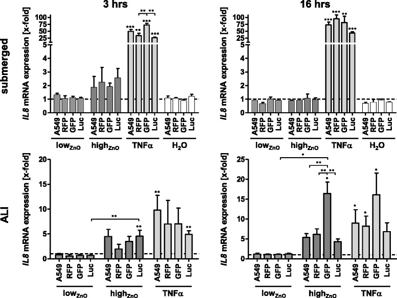
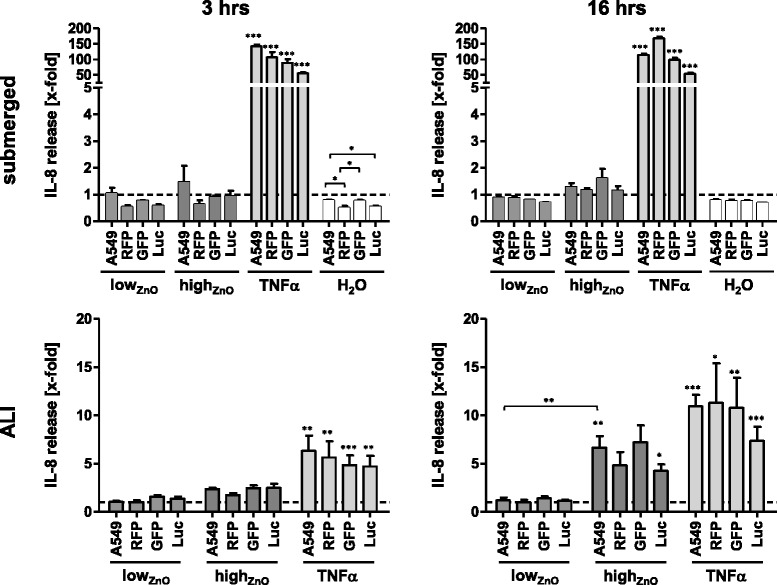
Methods: All cell lines were exposed to zinc oxide (ZnO) NPs at 0.6 and 6.2 μg/cm(2) for 3 and 16 hours under both submerged and ALI conditions. Following physicochemical characterization, the cytotoxic profile of the ZnO-NPs was determined for each exposure scenario. Expression of IL-8 from all cell types was analyzed at the promoter level and compared to the mRNA (qRT-PCR) and protein level (ELISA).
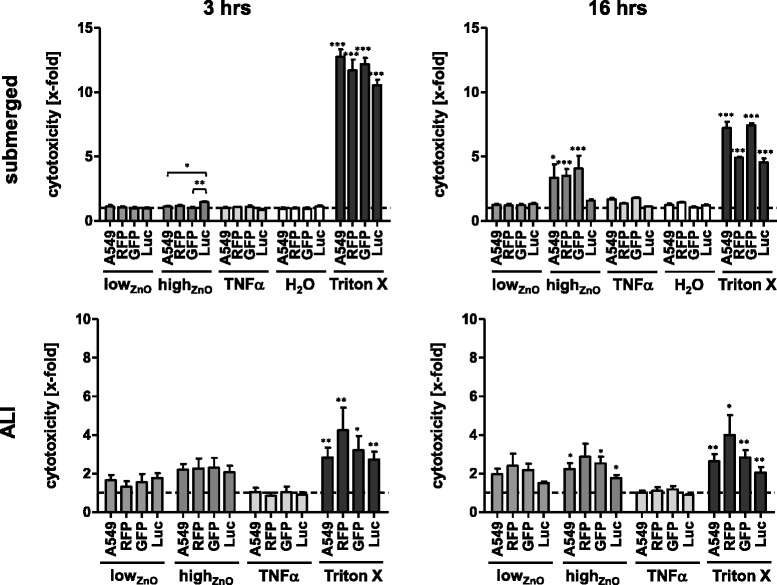
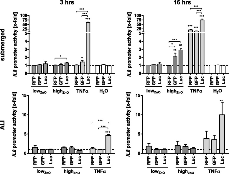
Results: In summary, each reporter cell line detected acute pro-inflammatory effects following ZnO exposure under each condition tested. The pIL8-Luc cell line was the most sensitive in terms of reporter signal strength and onset velocity following TNF-α treatment. Both pIL8-GFP and pIL8-RFP also showed a marked signal induction in response to TNF-α, although only after 16 hrs. In terms of ZnO-NP-induced cytotoxicity pIL8-RFP cells were the most affected, whilst the pIL8-Luc were found the least responsive.
Conclusions: In conclusion, the use of fluorescence-based reporter cell lines can provide a useful tool in screening the pro-inflammatory response following NP exposure in both submerged and ALI cell cultures.
Figures
References
-
- Westerink WM, Stevenson JC, Horbach GJ, Schoonen WG. The development of RAD51C, Cystatin A, p53 and Nrf2 luciferase-reporter assays in metabolically competent HepG2 cells for the assessment of mechanism-based genotoxicity and of oxidative stress in the early research phase of drug development. Mutat Res. 2010;696(1):21–40. doi: 10.1016/j.mrgentox.2009.12.007. - DOI - PubMed
Publication types
MeSH terms
Substances
LinkOut - more resources
Full Text Sources
Other Literature Sources
Miscellaneous
