. 2015 Oct 28;7(40):16571-7.
doi: 10.1039/c5nr03411f.

Hybrid upconversion nanomaterials for optogenetic neuronal control

Affiliations

Hybrid upconversion nanomaterials for optogenetic neuronal control

Shreyas Shah et al. Nanoscale. .

Abstract

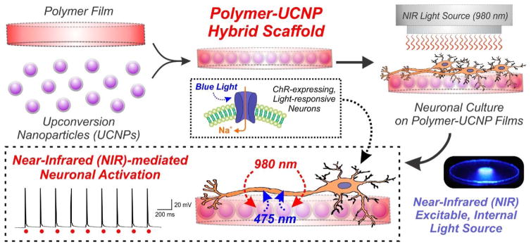
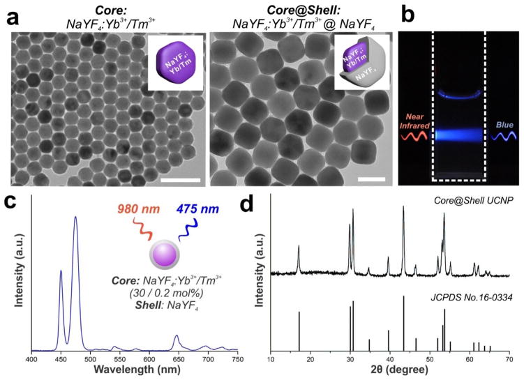
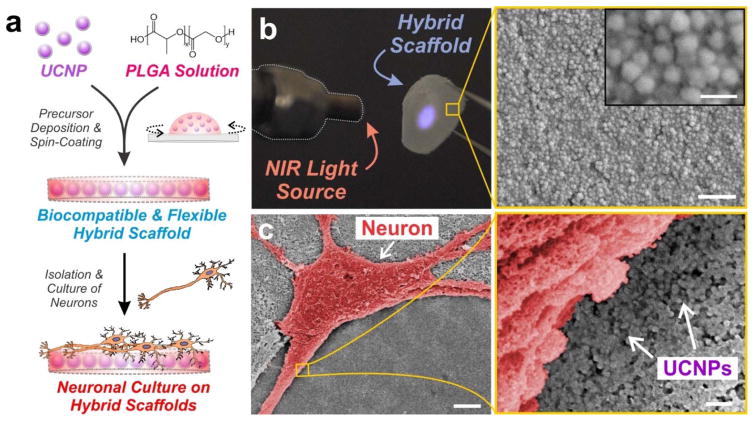
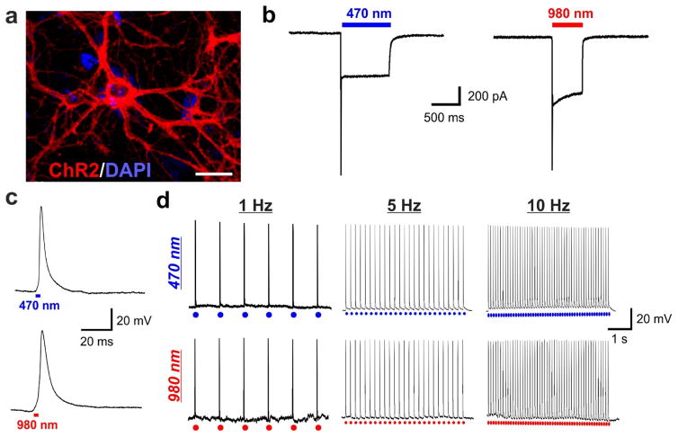
Nanotechnology-based approaches offer the chemical control required to develop precision tools suitable for applications in neuroscience. We report a novel approach employing hybrid upconversion nanomaterials, combined with the photoresponsive ion channel channelrhodopsin-2 (ChR2), to achieve near-infrared light (NIR)-mediated optogenetic control of neuronal activity. Current optogenetic methodologies rely on using visible light (e.g. 470 nm blue light), which tends to exhibit high scattering and low tissue penetration, to activate ChR2. In contrast, our approach enables the use of 980 nm NIR light, which addresses the short-comings of visible light as an excitation source. This was facilitated by embedding upconversion nanomaterials, which can convert NIR light to blue luminescence, into polymeric scaffolds. These hybrid nanomaterial scaffolds allowed for NIR-mediated neuronal stimulation, with comparable efficiency as that of 470 nm blue light. Our platform was optimized for NIR-mediated optogenetic control by balancing multiple physicochemical properties of the nanomaterial (e.g. size, morphology, structure, emission spectra, concentration), thus providing an early demonstration of rationally-designing nanomaterial-based strategies for advanced neural applications.


Figures

Figure 1
Figure 1. Schematic diagram depicting the generation and application of polymer-UCNP hybrid scaffolds for optogenetic neuronal activation
Upconversion nanoparticles (UCNPs) were embedded within polymeric films to form biocompatible hybrid scaffolds for neuronal culture. The UCNPs served as internally excitable light sources that convert NIR light into blue light, thus facilitating optogenetic activation of channelrhodopsin (ChR)-expressing neurons.
Figure 2
Figure 2. Monodisperse core-shell UCNPs with blue emission upon near-infrared excitation were synthesized
(a) Transmission electron microscopy (TEM) image of core NaYF4:Yb3+/Tm3+ (30 / 0.2 mol%) nanoparticles (left, scale bar: 100 nm) and core@shell NaYF4:Yb3+/Tm3+ (30 / 0.2 mol%)@NaYF4 UCNPs (right, scale bar: 50 nm). (b) Photographic image of a cuvette with a suspension of UCNPs under laser excitation at 980-nm. (c) Upconversion emission spectrum of core-shell UCNPs in hexane solution (1 mg/mL) under near-infrared light excitation at 980-nm. (d) Powder X-ray diffraction (PXRD) patterns of the core-shell UCNPs, showing all peaks to be well-indexed in accordance with the β-hexagonal-phase NaYF4 crystal structure (Joint Committee on Powder Diffraction Standards file No. 16-0334).
Figure 3
Figure 3. Blue-emitting UCNPs were embedded within thin polymer films of PLGA and cultured with hippocampal neurons
(a) Schematic depicting preparation of poly(lactic-co-glycolic acid) (PLGA)-embedded UCNP hybrid scaffolds for hippocampal neuronal cultures. (b) Photographic image of the flexible hybrid scaffold film. The blue luminescence under 980-nm laser excitation illustrates the encapsulation of UCNPs throughout the PLGA film. Scanning electron microscopy (SEM) image shows the distribution of the UCNPs within the PLGA film. Scale bar: 500 nm (bottom), 100 nm (inset). (c) Scanning electron microscopy (SEM) image of a hippocampal neuron (pseudo-colored red for contrast) cultured on the polymer-UCNP hybrid scaffolds at 14 DIV. Scale bar: 5 μm (left), 200 nm (right).
Figure 4
Figure 4. Channelrhodopsin-expressing neurons grown on polymer-UCNP films exhibit robust excitatory activation upon NIR-light illumination, comparable to blue LED illumination
(a) Hippocampal neurons expressing ChR2-tdTomato (red). Scale bar: 50 μm. (b) Representative traces of inward current flow in voltage-clamped neuron evoked by 470-nm light (5 mW) and 980-nm light (1 W). (c) Magnified action potential induced by 470-nm light (top) and 980-nm light (bottom). (d) Representative traces showing repetitive action potentials in a current-clamped hippocampal neuron evoked by 1 Hz, 5 Hz and 10 Hz train of light pulses from 470-nm light (top; 250 mW, 3 ms pulse width) and 980-nm light (bottom; 1 W, 3 ms pulse width).

References

    1. Alivisatos AP, Andrews AM, Boyden ES, Chun M, Church GM, Deisseroth K, Donoghue JP, Fraser SE, Lippincott-Schwartz J, Looger LL, Masmanidis S, McEuen PL, Nurmikko AV, Park H, Peterka DS, Reid C, Roukes ML, Scherer A, Schnitzer M, Sejnowski TJ, Shepard KL, Tsao D, Turrigiano G, Weiss PS, Xu C, Yuste R, Zhuang X. ACS Nano. 2013;7:1850–1866. - PMC - PubMed
    1. Boyden ES, Zhang F, Bamberg E, Nagel G, Deisseroth K. Nat Neurosci. 2005;8:1263–1268. - PubMed
    1. Zhang F, Gradinaru V, Adamantidis AR, Durand R, Airan RD, de Lecea L, Deisseroth K. Nat Protoc. 2010;5:439–456. - PMC - PubMed
    1. Tye KM, Deisseroth K. Nat Rev Neurosci. 2012;13:251–266. - PMC - PubMed
    1. Brieke C, Rohrbach F, Gottschalk A, Mayer G, Heckel A. Angew Chem Int Ed. 2012;51:8446–8476. - PubMed

Publication types

Substances