

Point-of-Care Ultrasound Assessment of Tropical Infectious Diseases--A Review of Applications and Perspectives
- PMID: 26416111
- PMCID: PMC4710450
- DOI: 10.4269/ajtmh.15-0421
Point-of-Care Ultrasound Assessment of Tropical Infectious Diseases--A Review of Applications and Perspectives
Abstract
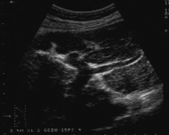
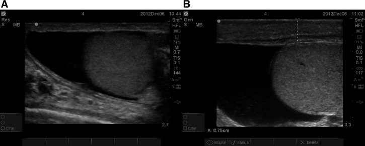
The development of good quality and affordable ultrasound machines has led to the establishment and implementation of numerous point-of-care ultrasound (POCUS) protocols in various medical disciplines. POCUS for major infectious diseases endemic in tropical regions has received less attention, despite its likely even more pronounced benefit for populations with limited access to imaging infrastructure. Focused assessment with sonography for HIV-associated TB (FASH) and echinococcosis (FASE) are the only two POCUS protocols for tropical infectious diseases, which have been formally investigated and which have been implemented in routine patient care today. This review collates the available evidence for FASH and FASE, and discusses sonographic experiences reported for urinary and intestinal schistosomiasis, lymphatic filariasis, viral hemorrhagic fevers, amebic liver abscess, and visceral leishmaniasis. Potential POCUS protocols are suggested and technical as well as training aspects in the context of resource-limited settings are reviewed. Using the focused approach for tropical infectious diseases will make ultrasound diagnosis available to patients who would otherwise have very limited or no access to medical imaging.
© The American Society of Tropical Medicine and Hygiene.
Figures

References
-
- Richter J, Hatz C, Haussinger D. Ultrasound in tropical and parasitic diseases. Lancet. 2003;362:900–902. - PubMed
-
- Moore CL, Copel JA. Point-of-care ultrasonography. N Engl J Med. 2011;364:749–757. - PubMed
-
- Hanscheid T, Rebelo M, Grobusch MP. Point-of-care tests: where is the point? Lancet. 2014;14:922. - PubMed
-
- Ma OJ, Mateer JR, Ogata M, Kefer MP, Wittmann D, Aprahamian C. Prospective analysis of a rapid trauma ultrasound examination performed by emergency physicians. J Trauma. 1995;38:879–885. - PubMed
-
- Mateer J, Plummer D, Heller M, Olson D, Jehle D, Overton D, Gussow L. Model curriculum for physician training in emergency ultrasonography. Ann Emerg Med. 1994;23:95–102. - PubMed
Publication types
MeSH terms
LinkOut - more resources
Full Text Sources
Other Literature Sources
Medical
