. 2015 Oct 1;526(7571):136-9.
doi: 10.1038/nature15254. Epub 2015 Sep 23.

Multiple mechanisms for CRISPR-Cas inhibition by anti-CRISPR proteins

Affiliations

Multiple mechanisms for CRISPR-Cas inhibition by anti-CRISPR proteins

Joseph Bondy-Denomy et al. Nature. .

Abstract

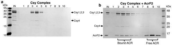
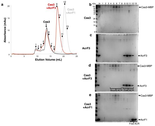
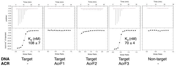
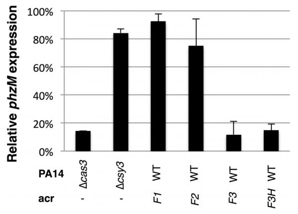
The battle for survival between bacteria and the viruses that infect them (phages) has led to the evolution of many bacterial defence systems and phage-encoded antagonists of these systems. Clustered regularly interspaced short palindromic repeats (CRISPR) and the CRISPR-associated (cas) genes comprise an adaptive immune system that is one of the most widespread means by which bacteria defend themselves against phages. We identified the first examples of proteins produced by phages that inhibit a CRISPR-Cas system. Here we performed biochemical and in vivo investigations of three of these anti-CRISPR proteins, and show that each inhibits CRISPR-Cas activity through a distinct mechanism. Two block the DNA-binding activity of the CRISPR-Cas complex, yet do this by interacting with different protein subunits, and using steric or non-steric modes of inhibition. The third anti-CRISPR protein operates by binding to the Cas3 helicase-nuclease and preventing its recruitment to the DNA-bound CRISPR-Cas complex. In vivo, this anti-CRISPR can convert the CRISPR-Cas system into a transcriptional repressor, providing the first example-to our knowledge-of modulation of CRISPR-Cas activity by a protein interactor. The diverse sequences and mechanisms of action of these anti-CRISPR proteins imply an independent evolution, and foreshadow the existence of other means by which proteins may alter CRISPR-Cas function.


Figures

Extended Data Figure 1
Extended Data Figure 1. AcrF2 interacts with the Csy complex
a, b, Purified Csy complex was fractionated by SEC alone (a) or in the presence of AcrF2 (b). Fractions were analysed on a silver nitrate stained SDS–PAGE gel. The input (IN) and fractions are shown.
Extended Data Figure 2
Extended Data Figure 2. AcrF3, not AcrF1, interacts with Cas3
a, Cas3 was fractionated by SEC alone or in the presence of AcrF3 or AcrF1. Overlays of plots of elution volume versus optical density at 280 nm of the column eluates are shown. The numbers represent the fractions that were selected for analysis. b–e, Silver nitrate stained SDS–PAGE gels are shown from SEC experiments with Cas3 (b), AcrF3 (c), Cas3 with AcrF3 (d) or Cas3 with AcrF1 (e). The sample that was loaded onto the SEC column is shown as input (In) and fractions from the same elution positions are indicated numerically. AcrF3 is seen eluting in fractions 4–8 only in the presence of Cas3. There is also a visible shift in the Cas3 elution profile in the presence of AcrF3 but not AcrF1 (fractions 3–5).
Extended Data Figure 3
Extended Data Figure 3. AcrF1 and AcrF2 prevent target recognition by the Csy complex
Isothermal titration calorimetry (ITC) assays showing the Csy complex binding to an 8-nucleotide ssDNA target that comprises the seed region. No binding is observed in the presence of AcrF1, AcrF2 or with a non-target (the reverse complement sequence of the target) ssDNA substrate. A representative run is shown for each condition with the dissociation constant (Kd) value and error of fit from that particular run. Over multiple runs (n = 6) with the Csy complex binding to the ssDNA ligand, the average Kd value was 90 nM ± 37.
Extended Data Figure 4
Extended Data Figure 4. Expression of phzM is repressed by the Csy complex
The Csy complex was targeted to the promoter of the gene phzM, and repression efficiency was assayed by RT–qPCR (see Methods). The per cent repression of phzM in the indicated strains expressing a phzM-targeting crRNA relative to wild-type (WT) PA14 with an empty plasmid is shown. All values were normalized to rpsL, a gene encoding a ribosomal protein. Means ± s.d. are shown.
Extended Data Figure 5
Extended Data Figure 5. AcrF4 interacts with the Csy complex
Untagged AcrF4 was expressed in E. coli BL21 cells and a crude lysate of these cells was mixed with the Csy complex bound to Ni-NTA beads via a 6×His tag on Csy3. a, The flow through (FT), wash 1 (W1), and two elution fractions (E1, E2) from the Ni-NTA column are shown, as well as a comparison to pure Csy complex. b, The Ni-NTA elution fractions were fractionated by SEC, demonstrating a stable interaction between the Csy complex and AcrF4. The input (In) lane shows the sample that was loaded on the SEC column and numbered fractions are analysed on SDS–PAGE gels.
Extended Data Figure 6
Extended Data Figure 6. AcrF1 and AcrF2 bind the Csy complex at distinct locations
a, Purified Csy1–Csy2 heterodimer with an MBP and 6×His tag fused to Csy1 was fractionated by SEC in the presence or absence of AcrF1 (boxes indicate the Csy1–Csy2 peak). b, Purified MBP/6×His-tagged Csy3 was fractionated in the presence or absence of AcrF2. These are complementary experiments to those seen in Fig. 3b and c, respectively. Input (In) and selected fractions are shown on SDS–PAGE gels. c, AcrF1 and AcrF2 were incubated with the Csy complex singly or in combination. Asterisks designate which anti-CRISPR was added first to the reactions containing both anti-CRISPR proteins. The addition order did not affect the result since there is no competition for binding sites between these two anti-CRISPR proteins. After incubation, each mixture was fractionated by SEC and the peak Csy complex fraction is shown on an SDS–PAGE gel. In each experiment the anti-CRISPR proteins are in excess relative to the Csy complex.
Extended Data Figure 7
Extended Data Figure 7. AcrF1 and AcrF2 interact with an RNase-A-treated Csy complex
a, The Csy complex was treated with a low concentration (600 nM, +) of RNase A or a high concentration of RNase A (70 μM, ++). This mixture was fractionated by SEC, revealing Csy4 dissociation at the higher RNase A concentration. Pre-treatment of the Csy complex with RNase A, with the subsequent addition of AcrF1 or AcrF2 followed by SEC fractionation was then conducted. Peak Csy complex fractions are shown on an SDS–PAGE gel. b, A TBE-urea denaturing gel is shown, stained with SYBR gold, showing the native crRNA in the Csy complex and the protected fragments remaining after 70 μM RNase A treatment. c, Quantification of Coomassie blue stained gels from three independent preparations of the respective proteins is shown. Anti-CRISPR proteins bound with unaltered stoichiometry to RNase-A-pre-treated Csy complexes. Error bars represent s.d.
Extended Data Figure 8
Extended Data Figure 8. Twofold dilutions used to quantify anti-CRISPR binding stoichiometry
Csy complexes with crRNA molecules possessing spacers of differing lengths (16, 32, or 48 nucleotides) were purified and fractionated by SEC in the presence of AcrF1. A representative Coomassie blue stained SDS–PAGE gel is shown, with twofold dilutions of the peak fraction containing the Csy complex and co-eluting AcrF1. Arrows on the bottom of the gel indicate comparable dilutions based on the levels of Csy1. Note the increasing abundance of Csy3 and AcrF1. b, Lanes with arrows from the gel in a are shown next to each other for comparison.
Extended Data Figure 9
Extended Data Figure 9. dsDNA binds to the Csy complex after SEC fractionation
a, The same samples from Fig. 4a were run on a denaturing TBE-urea gel, stained with SYBR gold, to reveal the crRNA (two species are apparent), and the Csy-complex-bound 50 bp dsDNA. In these experiments, DNA was prebound to the Csy complex, and AcrF1 or AcrF2 were subsequently added to the DNA-saturated Csy complex. This mixture was then fractionated by SEC and the Csy-complex-containing peak fractions were analysed. b, A schematic showing the crRNA sequence with repeat-derived regions shown in black and the variable 32-nucleotide spacer region in red. The seed-interacting region that is critical for target recognition (nucleotides 1–5, 7, 8) is in bold. DNA oligonucleotides used in this study are shown, with labels ‘A’, ‘B’ and ‘C’ corresponding to the targets shown in Fig. 4c. The 8-nucleotide ssDNA substrate was used in ITC experiments (Extended Data Fig. 3), and the 50 bp dsDNA in EMSAs (Figs 1d and 4b).
Figure 1
Figure 1. Anti-CRISPR proteins inhibit CRISPR–Cas function by directly interacting with the Csy complex or Cas3
a, b, Purified Csy complex was incubated with purified AcrF1 (a) or AcrF3 (b) and the mixture was fractionated by SEC. Fractions were analysed by SDS–polyacrylamide gel electrophoresis (SDS–PAGE) and are numbered according to their elution position (see Extended Data Fig. 1 for SEC of the Csy complex alone or with AcrF2). The purified Csy complex or anti-CRISPR (ACR) are shown in the second (Csy) and last (ACR) lanes, respectively. c, Purified Cas3 was incubated with (right) or without (left) AcrF3 and fractionated by SEC. The eluting fractions were analysed by SDS–PAGE as described earlier. The input (In) lanes show the protein mixture that was loaded onto the SEC column. MBP, maltose-binding protein. d, dsDNA binding by the Csy complex was assayed using an EMSA. Csy complex was present in all reactions except for lanes 1 and 6. Other components added to each reaction are designated above the lanes. In the lanes coloured red and blue, the designated components were premixed before the addition of DNA. ATP was added to the Csy–DNA–Cas3 reaction either before the addition of Cas3 (lanes 11, 12) or after (lane 13). The supershifted species resulting from Cas3 addition did not migrate into the gel upon prolonged electrophoresis, but it is dissociated by the addition of ATP (lane 13), demonstrating that the supershift is not caused by aggregated inactive protein.
Figure 2
Figure 2. Anti-CRISPR proteins interact with Cas proteins in vivo
a, The phzM promoter was targeted by a plasmid-encoded crRNA in P. aeruginosa. The production of pyocyanin was quantified in different PA14 mutant backgrounds (Δcas3, Δcsy3 or ΔphzM) or during the expression of the indicated anti-CRISPR from a prophage. The amount of pyocyanin produced in the presence of a plasmid producing the crRNA is shown as a percentage of the same strain with the empty plasmid vector. An average of three independent experiments is shown with error bars representing the standard deviation (s.d.). Representative pictures of cultures are shown. The pyocyanin ratio for the ΔphzM mutant was derived by comparing it to the value for the Δcsy3 mutant. The prophage expressing acrF3 also encoded another anti-CRISPR, the functional mechanism of which is not known. To bolster our conclusions pertaining to acrF3, we also tested a prophage that expresses an 86% identical homologue of acrF3, designated acrF3H, and no other anti-CRISPR. WT, wild type. b, Lysates of phages expressing the indicated anti-CRISPR proteins were spotted in tenfold serial dilutions on bacterial lawns of wild-type P. aeruginosa PA14 (top) or the same strain bearing a plasmid that overexpresses the Csy subunits (bottom). These phages would be targeted by the CRISPR–Cas system in the absence of anti-CRISPR activity.
Figure 3
Figure 3. AcrF1 and AcrF2 bind distinct Csy complex subunits
a, A schematic of the crRNA showing the repeat-derived regions of the crRNA (black) and the 32-nucleotide (nt) spacer region (red). The coloured circles represent the Csy1–4 subunits. b, c, Purified 6×His/MBP-tagged Csy1–Csy2 heterodimer (b) or Csy3 (c) was fractionated by SEC in the presence (right) or absence (left) of the indicated anti-CRISPR proteins. The SEC fractions were analysed by SDS–PAGE. The ‘In’ lanes show the protein mixture that was loaded onto the SEC column and fractions are numbered. d, Purified Csy complexes with 16-, 32-, or 48-nucleotide crRNA spacer regions were bound to AcrF1 or AcrF2 and fractionated by SEC. The stoichiometry of the bound anti-CRISPR proteins was quantified through densitometry of the Coomassie blue stained gels. An average of three independent experiments is shown with error bars representing s.d.
Figure 4
Figure 4. Two anti-CRISPR proteins inhibit target recognition via unique mechanisms
a, EMSA experiments were used to assay binding of the Csy complex to three different ssDNA oligonucleotides (labelled A, B and C) that are complementary to different regions of the crRNA spacer as shown in the schematic (see Extended Data Fig. 9b). Where noted, the Csy complex was pre-incubated with the indicated anti-CRISPR. b, c, Apo–Csy complex (AC) or DNA-bound Csy complex (DC) was incubated with AcrF1 or AcrF2. b, This mixture was fractionated by SEC and fractions were visualized by SDS–PAGE. c, An EMSA experiment is shown with binding to dsDNA in the same experimental setup as in b. d, A model summarizing anti-CRISPR mechanisms. Arrows indicate the steps of the uninhibited CRISPR–Cas interference pathway. Numbers in the Csy complex indicate the Csy subunits. The lines with flat ends indicate the step in the CRISPR–Cas pathway blocked by each anti-CRISPR. The manner in which each anti-CRISPR binds to CRISPR–Cas components is also shown. AcrF1 makes the whole crRNA inaccessible while AcrF2 occludes the 5′ end.

Comment in

References

    1. Barrangou R, et al. CRISPR provides acquired resistance against viruses in prokaryotes. Science. 2007;315:1709–1712. - PubMed
    1. Makarova KS, et al. Evolution and classification of the CRISPR-Cas systems. Nature Rev. Microbiol. 2011;9:467–477. - PMC - PubMed
    1. Jore MM, Brouns SJJ, van der Oost J. RNA in defense: CRISPRs protect prokaryotes against mobile genetic elements. Cold Spring Harb. Perspect. Biol. 2012;4:a003657. - PMC - PubMed
    1. Bondy-Denomy J, Pawluk A, Maxwell KL, Davidson AR. Bacteriophage genes that inactivate the CRISPR/Cas bacterial immune system. Nature. 2013;493:429–432. - PMC - PubMed
    1. van der Oost J, Westra ER, Jackson RN, Wiedenheft B. Unravelling the structural and mechanistic basis of CRISPR–Cas systems. Nature Rev. Microbiol. 2014;12:479–492. - PMC - PubMed

Publication types

MeSH terms