. 2015 Dec;35(24):4110-20.
doi: 10.1128/MCB.00354-15. Epub 2015 Sep 28.

PAK1 and CtBP1 Regulate the Coupling of Neuronal Activity to Muscle Chromatin and Gene Expression

Affiliations

PAK1 and CtBP1 Regulate the Coupling of Neuronal Activity to Muscle Chromatin and Gene Expression

Jean-Luc Thomas et al. Mol Cell Biol. 2015 Dec.

Abstract

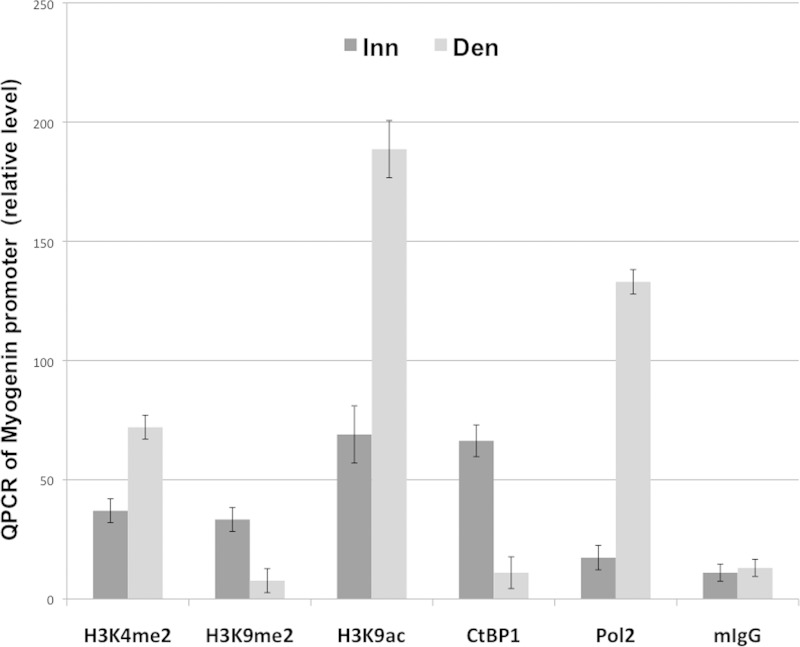
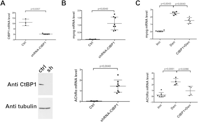
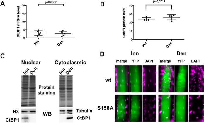
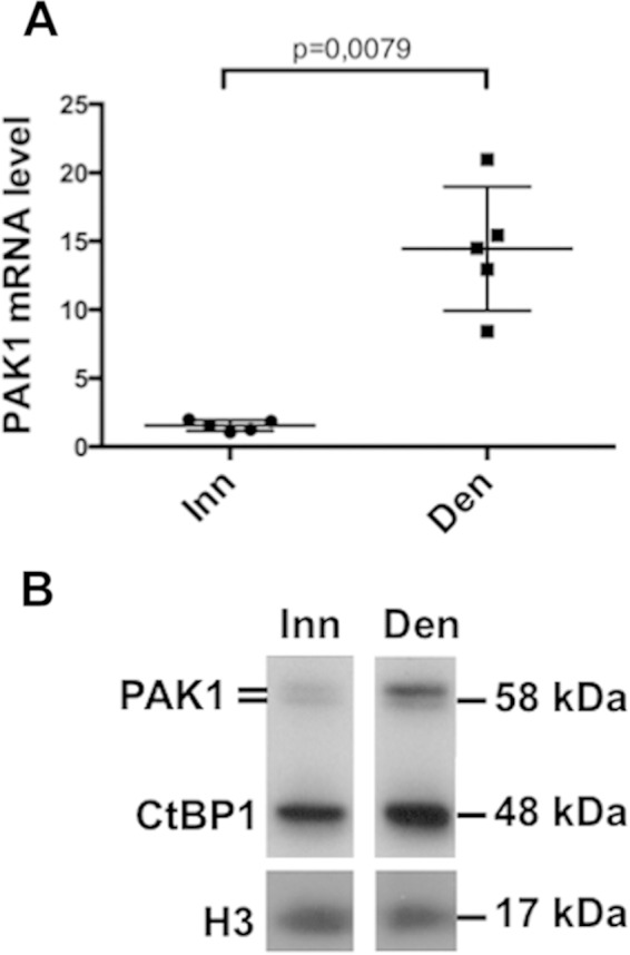
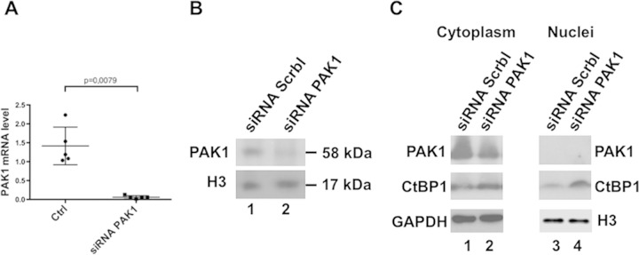
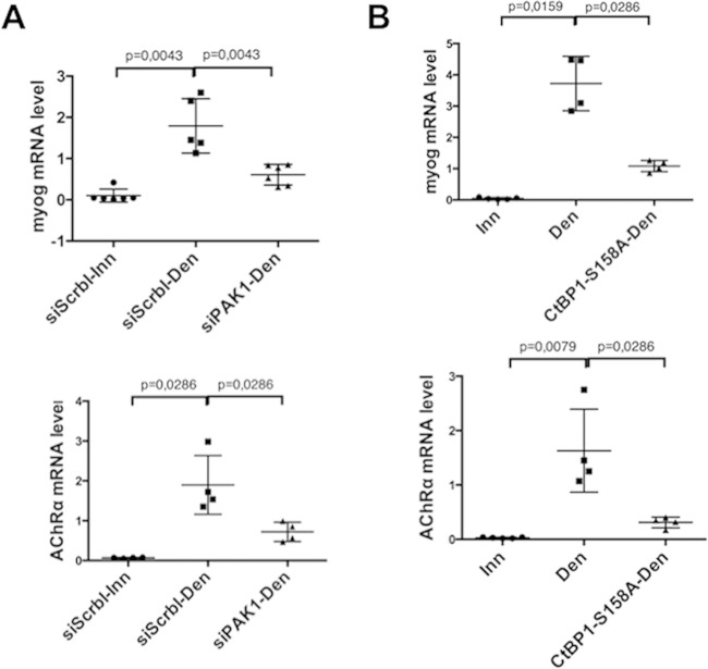
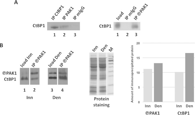
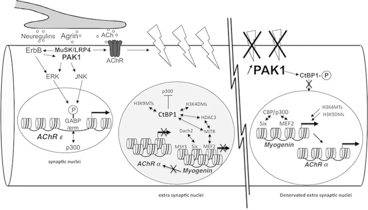
Acetylcholine receptor (AChR) expression in innervated muscle is limited to the synaptic region. Neuron-induced electrical activity participates in this compartmentalization by promoting the repression of AChR expression in the extrasynaptic regions. Here, we show that the corepressor CtBP1 (C-terminal binding protein 1) is present on the myogenin promoter together with repressive histone marks. shRNA-mediated downregulation of CtBP1 expression is sufficient to derepress myogenin and AChR expression in innervated muscle. Upon denervation, CtBP1 is displaced from the myogenin promoter and relocates to the cytoplasm, while repressive histone marks are replaced by activating ones concomitantly to the activation of myogenin expression. We also observed that upon denervation the p21-activated kinase 1 (PAK1) expression is upregulated, suggesting that phosphorylation by PAK1 may be involved in the relocation of CtBP1. Indeed, preventing CtBP1 Ser158 phosphorylation induces CtBP1 accumulation in the nuclei and abrogates the activation of myogenin and AChR expression. Altogether, these findings reveal a molecular mechanism to account for the coordinated control of chromatin modifications and muscle gene expression by presynaptic neurons via a PAK1/CtBP1 pathway.


Figures

FIG 1
FIG 1
ChIP analysis of the myogenin promoter. Innervated or denervated muscle extracts were used for ChIP experiments with antibodies against H3K4me2, H3K9me2, H3ac, CtBP1, the RNA polymerase 2 largest subunit (Pol2), or mouse IgG (mIgG). Immunoprecipitated DNA was amplified by Q-PCR with primers spanning the myogenin promoter (see Materials and Methods). Inn, innervated; Den, denervated.
FIG 2
FIG 2
CtBP1 regulates myogenin and AChrα expression. Each point corresponds to the analysis of a different TA muscle. The central bars, representing the means, and standard deviations are indicated. In each panel, the first lane shows the initial level of mRNA in innervated muscle. (A) Effects of CtBP1 shRNA on CtBP1 mRNA level (upper panel) or CtBP1 protein level (lower panel). (B) Effects of CtBP1 shRNA on myogenin mRNA level (upper panel) or AChRα mRNA level (lower panel). (C) Effect of overexpressed recombinant CtBP1 in denervated muscle on myogenin (upper panel) or AChRα (lower panel) mRNA levels. In panels A and B, Ctrl corresponds to the control with scramble shRNA. In panel C, Inn and Den correspond, respectively, to the control of innervated and denervated muscles treated with the empty vector.
FIG 3
FIG 3
Innervation regulates the nuclear localization of CtBP1. (A) CtBP1 mRNAs were quantified by quantitative RT-PCR in innervated or denervated muscles extracts. (B) CtBP1 protein levels in innervated or denervated muscle extracts quantified by Western blotting. (C) Western blot of CtBP1 in cytoplasmic and nuclear extracts of innervated or denervated muscle. Identical amounts of protein extracts, as shown by protein staining, were separated using 10% PAGE, and the protein contents were analyzed by Western blotting with CtBP1 antibody and either a histone H3 antibody (for nuclear extracts) or a tubulin antibody (for cytoplasmic extracts). (D) Detection of labeled nuclei after electroporation of a CtBP1-YFP plasmid (wt) or a CtBP1-S158A-YFP plasmid (S158A) in innervated (Inn) or denervated (Den) muscle. YFP detection, DAPI, and merge images are indicated. One representative fiber is shown in each case.
FIG 4
FIG 4
PAK1 level is regulated by innervation. The levels of PAK1 mRNA detected by RT-PCR (A) and the PAK1 protein detected by Western blotting (B) in innervated and denervated TA muscles were determined.
FIG 5
FIG 5
Inhibition of PAK1 expression induces an increase in nuclear CtBP1 level. (A) Levels of PAK1 mRNA detected by quantitative RT-PCR after electroporation of PAK1 siRNA (right) or scramble siRNA (left) in innervated muscle. (B) Levels of PAK1 protein detected by Western blotting in a total TA muscle extract after coelectroporation of mCherry-Nuc with scramble siRNA in denervated muscle (lane 1) or coelectroporation of mCherry-Nuc with PAK1 siRNA in denervated muscle (lane 2). (C) Effect of the PAK1 siRNA on the levels of CtBP1 protein detected by Western blotting in TA cytoplasmic (lanes 1 and 2) or nuclear (lanes 3 and 4) extracts from denervated muscle electroporated with Scramble siRNA (lanes 1 to 3) or with PAK1 siRNA (lanes 2 to 4).
FIG 6
FIG 6
Effects of PAK1 siRNA (A) or CtBP1-S158A mutant (B) on the levels of myogenin (upper panel) and AChRα (lower panel) mRNA in denervated tibialis anterior muscle, as detected by RT-PCR. In each panel the left column shows the data for a control (innervated muscle electroporated with scramble siRNA in panel A; innervated muscle in panel B), the middle column shows the effect of denervation (including electroporation with Scramble siRNA in panel A), and the right column shows the effect of PAK1 siRNA electroporation in denervated muscle (A) or the effect of CtBP1-S158A overexpression in denervated muscle (B).
FIG 7
FIG 7
Interaction between CtBP1 and PAK1/P-PAK1. (A) The left panel shows Western blot detection of CtBP1 immunoprecipitated by CtBP1 antibody (lane 1), PAK1 antibody (lane 2), or mouse IgG antibody (lane 3); the right panel shows Western blot detection of CtBP1 in tibialis anterior extract (lane 1) and immunoprecipitated by mouse IgG antibody (lane 2) or P-PAK1 antibody (lane 3). (B) Comparison of CtBP1 and P-PAK1 immunoprecipitation by P-PAK1 antibody in innervated versus denervated muscle extracts. The left panel shows Western blot detection of CtBP1 and P-PAK1 in the input extracts (lanes 1 and 3) and in the immunoprecipitated fractions (lanes 2 and 4) from either innervated (Inn: lanes 1 to 2) or denervated (Den, lanes 3 to 4) extracts. In the middle panel, Coomassie blue staining shows equal loading of the input extracts. In the right panel, histograms based on scanning of the Western blot bands show that more CtBP1 is immunoprecipitated from the denervated extract than from the innervated extract, while a similar amount of P-PAK1 was immunoprecipitated. Circled P, phosphorylation.
FIG 8
FIG 8
PAK1 and CtBP1 mediate the action of neural inputs on gene expression in muscle. Upon binding to LRP4, agrin induces the aggregation of synaptic proteins, including AChR and ErbB receptors. It also activates gene expression via the ERK and JNK intracellular signaling pathways that phosphorylate and activate Ets transcription factors. PAK1 is activated by the neural factors agrin and neuregulins via MuSK/LRP4 and ErbB receptors, respectively, and is involved in the cytoskeletal network organization. In extrasynaptic nuclei, CtBP1 is recruited on the promoter of the myogenin gene, where it participates in repression by inhibiting the p300/CBP histone acetyltransferase and by recruiting histone deacetylases, H3K4 demethylases, and H3K9 methyltransferases. In denervated muscle fibers, PAK1 expression is significantly activated in extrasynaptic regions. Increased PAK1 levels induce CtBP1 phosphorylation (indicated by a “circled P” in the figure) and cytoplasmic localization, thereby allowing the recruitment of H3K4 methyltransferases and H3K9 demethylases on the myogenin promoter, as well as the coactivation of Six and MEF2 by CBP/p300. Myogenin then activates the expression of the AChR α, β, δ, and γ subunit genes.

References

    1. Burden SJ. 2011. SnapShot: neuromuscular junction. Cell 144:826–826.e1. doi: 10.1016/j.cell.2011.02.037. - DOI - PubMed
    1. Wu H, Xiong WC, Mei L. 2010. To build a synapse: signaling pathways in neuromuscular junction assembly. Development 137:1017–1033. doi: 10.1242/dev.038711. - DOI - PMC - PubMed
    1. Luo ZG, Wang Q, Zhou JZ, Wang J, Luo Z, Liu M, He X, Wynshaw-Boris A, Xiong WC, Lu B, Mei L. 2002. Regulation of AChR clustering by Dishevelled interacting with MuSK and PAK1. Neuron 35:489–505. doi: 10.1016/S0896-6273(02)00783-3. - DOI - PubMed
    1. Ravel-Chapuis A, Vandromme M, Thomas JL, Schaeffer L. 2007. Postsynaptic chromatin is under neural control at the neuromuscular junction. EMBO J 26:1117–1128. doi: 10.1038/sj.emboj.7601572. - DOI - PMC - PubMed
    1. Mejat A, Ramond F, Bassel-Duby R, Khochbin S, Olson EN, Schaeffer L. 2005. Histone deacetylase 9 couples neuronal activity to muscle chromatin acetylation and gene expression. Nat Neurosci 8:313–321. doi: 10.1038/nn1408. - DOI - PubMed

Publication types

MeSH terms