. 2015 Dec;83(12):4682-92.
doi: 10.1128/IAI.00775-15. Epub 2015 Sep 28.

Important contribution of the novel locus comEB to extracellular DNA-dependent Staphylococcus lugdunensis biofilm formation

Affiliations

Important contribution of the novel locus comEB to extracellular DNA-dependent Staphylococcus lugdunensis biofilm formation

Nithya Babu Rajendran et al. Infect Immun. 2015 Dec.

Abstract

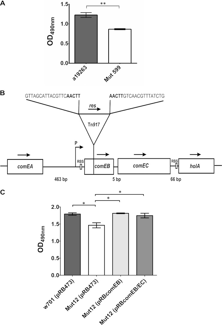
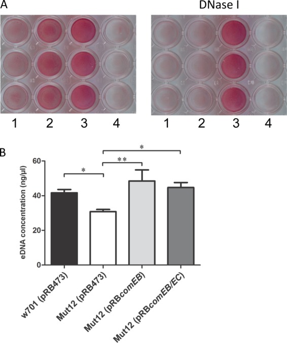
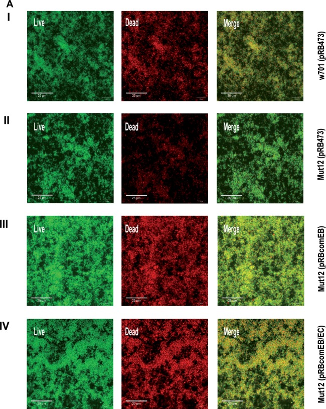
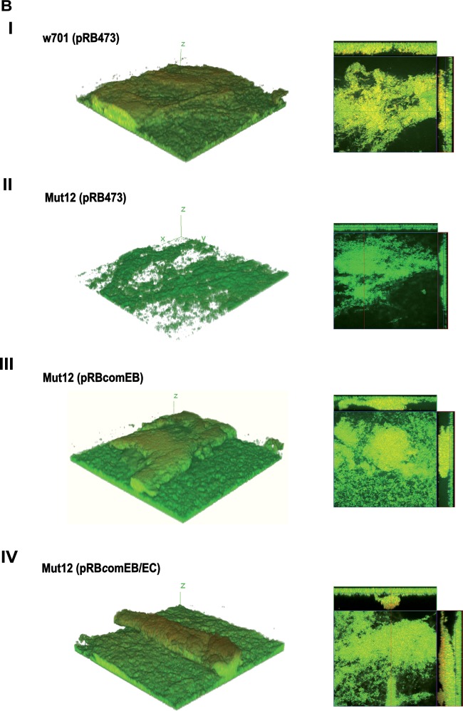
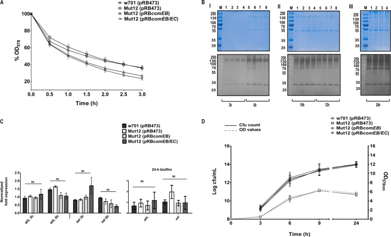
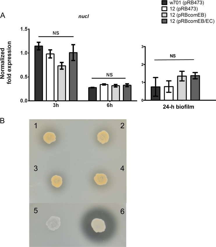
The coagulase-negative species Staphylococcus lugdunensis is an emerging cause of serious and potentially life-threatening infections, such as infective endocarditis. The pathogenesis of these infections is characterized by the ability of S. lugdunensis to form biofilms on either biotic or abiotic surfaces. To elucidate the genetic basis of biofilm formation in S. lugdunensis, we performed transposon (Tn917) mutagenesis. One mutant had a significantly reduced biofilm-forming capacity and carried a Tn917 insertion within the competence gene comEB. Site-directed mutagenesis and subsequent complementation with a functional copy of comEB verified the importance of comEB in biofilm formation. In several bacterial species, natural competence stimulates DNA release via lysis-dependent or -independent mechanisms. Extracellular DNA (eDNA) has been demonstrated to be an important structural component of many bacterial biofilms. Therefore, we quantified the eDNA in the biofilms and found diminished eDNA amounts in the comEB mutant biofilm. High-resolution images and three-dimensional data obtained via confocal laser scanning microscopy (CSLM) visualized the impact of the comEB mutation on biofilm integrity. The comEB mutant did not show reduced expression of autolysin genes, decreased autolytic activities, or increased cell viability, suggesting a cell lysis-independent mechanism of DNA release. Furthermore, reduced amounts of eDNA in the comEB mutant biofilms did not result from elevated levels or activity of the S. lugdunensis thermonuclease NucI. In conclusion, we defined here, for the first time, a role for the competence gene comEB in staphylococcal biofilm formation. Our findings indicate that comEB stimulates biofilm formation via a lysis-independent mechanism of DNA release.


Figures

FIG 1
FIG 1
comEB is involved in biofilm formation. (A) Quantitative analysis of 24-h biofilms produced by the Tn917 mutant Mut599 and its wild type. The OD490 values ± SEM are the averages from three independent experiments. **, P ≤ 0.01. (B) The Tn917 insertion site in Mut599 is located 64 bp downstream of the comEB start codon, which is part of a putative operon containing the comEB, comEC, and holA genes. The arrows mark the direction of transcription. P, putative promoter; RBS, ribosomal binding site; res, resolvase gene. The nucleotide sequences correspond to the regions flanking the transposon, with the nucleotides in boldface representing the characteristic 5-bp duplication. comEB, comEC, and holA putatively are transcribed from the common promoter upstream of comEB. (C) Quantitative analysis of 24-h biofilms produced by the clinical isolate S. lugdunensis w701 harboring the empty vector pRB473, its site-directed comEB mutant Mut12 containing pRB473, and the complemented mutants. For complementation, the vector pRB473 carrying comEB-comEC (pRBcomEB/EC) or comEB alone (pRBcomEB) was introduced into Mut12. The OD490 values ± SEM are averages from three independent assays. *, P ≤ 0.05. The data were analyzed with one-way ANOVA and a Bonferroni's posttest.
FIG 2
FIG 2
S. lugdunensis biofilm formation is eDNA dependent and comEB modulates eDNA levels in S. lugdunensis biofilms. (A) Biofilm formation in TSB in polystyrene microtiter plates in the presence (0.1 mg/ml) (right) or absence (left) of DNase I. Biofilms were stained with safranin. Lanes: 1, S. lugdunensis a19263; 2, S. lugdunensis w701; 3, S. epidermidis RP62A; 4, S. carnosus TM300. (B) Quantification of eDNA in biofilms. eDNA from 24-h biofilms was isolated and quantified by UV-Vis spectrophotometry. Mut12(pRB473) contained significantly less eDNA in the biofilm than its wild type, w701(pRB473), and the complemented mutants Mut12(pRBcomEB) and Mut12(pRBcomEB/EC). The values represented here are the averages from three independent experiments, and the error bars represent SEM. *, P ≤ 0.05; **, P ≤ 0.01. Statistical analysis was performed with one-way ANOVA and a Bonferroni's posttest.
FIG 3
FIG 3
CLSM analysis of biofilm. (A) To visualize eDNA, mature 24-h biofilms grown on coverslips were stained with SYTO9 (live; green fluorescent) and PI (dead; red fluorescent) and are presented as maximum intensity projections. Colocalization occurs when live cells are closely associated with dead cells or are covered with eDNA, appearing yellow in the merged image (Merge). (B) To obtain 3D data, 48-h biofilms grown on coverslips were stained with the live/dead staining kit and 3D reconstructions were generated with z-stack images obtained with a z-slice interval of 1 μM. The images are representative of three independent experiments.
FIG 3
FIG 3
CLSM analysis of biofilm. (A) To visualize eDNA, mature 24-h biofilms grown on coverslips were stained with SYTO9 (live; green fluorescent) and PI (dead; red fluorescent) and are presented as maximum intensity projections. Colocalization occurs when live cells are closely associated with dead cells or are covered with eDNA, appearing yellow in the merged image (Merge). (B) To obtain 3D data, 48-h biofilms grown on coverslips were stained with the live/dead staining kit and 3D reconstructions were generated with z-stack images obtained with a z-slice interval of 1 μM. The images are representative of three independent experiments.
FIG 4
FIG 4
comEB does not impact the expression of autolytic activities. (A) Mid-log-phase cells were treated with Triton X-100, and autolysis was monitored by the drop in OD578. The OD values are expressed in percentages, with the initial OD set to 100%. n = 3. Error bars indicate SEM. (B) Surface-associated proteins prepared from bacterial strains that were harvested at different time points of growth (3 h, 6 h [I], 10 h, 12 h [II], and 24 h [III]) were separated by SDS-PAGE on SDS gels and corresponding zymogram gels. Zymogram gels contained heat-inactivated S. lugdunensis cells in the separation gel. Bacteriolytic activities were observed as clear zones after overnight incubation in buffer. (Upper) SDS gels; (lower) corresponding zymogram gels. Molecular masses (in kilodaltons) of marker proteins (M) are indicated on the left. Lanes 1 and 5, w701(pRB473); 2 and 6, Mut12(pRB473); 3 and 7, Mut12(pRBcomEB); 4 and 8, Mut12(pRBcomEB/EC). SDS and zymogram gels are representative of three independent experiments. (C) The expression of the S. lugdunensis homologs of two major staphylococcal autolysin genes, atlL and aal, were analyzed by real-time PCR from cultures grown to mid-logarithmic (3 h) or stationary phase (6 h) or in 24-h biofilms. The values represent the averages from three independent experiments, and the error bars represent the SEM. The data were analyzed with one-way ANOVA and a Bonferroni's posttest. (D) Growth was initiated at a starting OD578 of 0.05 in TSB (37°C, 160 rpm) and monitored. At the indicated time points, the cultures were sampled, serially diluted, and plated on blood agar. Resulting colonies were counted after 24 h of incubation. The viability was expressed as log CFU/ml. n = 3. Error bars indicate SEM. NS, not significant.
FIG 5
FIG 5
Expression (A) and activity (B) of the thermonuclease NucI is unchanged. (A) nucI gene expression was measured during the mid-logarithmic (3 h) and stationary (6 h) phases of growth or in 24-h-grown biofilms. The mRNA levels were quantified by real-time PCR and normalized against the relative quantities of the aroE and gyrB housekeeping gene transcripts. The data were analyzed with one-way ANOVA and a Bonferroni's posttest. (B) NucI activity, observed as clearing zones around bacterial growth on DNase agar, was similar among wild-type w701(pRB473) (2), Mut12(pRB473) (1), and the complemented mutants Mut12(pRBcomEB) (3) and Mut12(pRBcomEB/EC) (4). S. aureus SA113(pCU1) (6) and S. epidermidis O-47(pCU1) (5) served as positive and negative controls, respectively.

References

    1. Freney J, Brun Y, Bes M, Meugnier H, Grimont F, Grimont PAD, Nervi C, Fleurette J. 1988. Staphylococcus lugdunensis sp. nov. and Staphylococcus schleiferi sp. nov., two species from human clinical specimens. Int J Syst Bacteriol 38:168–172. doi:10.1099/00207713-38-2-168. - DOI
    1. Ravaioli S, Selan L, Visai L, Pirini V, Campoccia D, Maso A, Speziale P, Montanaro L, Arciola CR. 2012. Staphylococcus lugdunensis, an aggressive coagulase-negative pathogen not to be underestimated. Int J Artif Organs 35:742–753. - PubMed
    1. Becker K, Heilmann C, Peters G. 2014. Coagulase-negative staphylococci. Clin Microbiol Rev 27:870–926. doi:10.1128/CMR.00109-13. - DOI - PMC - PubMed
    1. Anguera I, Del Rio A, Miro JM, Matinez-Lacasa X, Marco F, Guma JR, Quaglio G, Claramonte X, Moreno A, Mestres CA, Mauri E, Azqueta M, Benito N, Garcia-de la Maria C, Almela M, Jimenez-Exposito MJ, Sued O, De Lazzari E, Gatell JM. 2005. Staphylococcus lugdunensis infective endocarditis: description of 10 cases and analysis of native valve, prosthetic valve, and pacemaker lead endocarditis clinical profiles. Heart 91:e10. doi:10.1136/hrt.2004.040659. - DOI - PMC - PubMed
    1. Böcher S, Tonning B, Skov RL, Prag J. 2009. Staphylococcus lugdunensis, a common cause of skin and soft tissue infections in the community. J Clin Microbiol 47:946–950. doi:10.1128/JCM.01024-08. - DOI - PMC - PubMed

Publication types

MeSH terms

Associated data

LinkOut - more resources