Review
. 2015 Dec 14;36(47):3320-31.
doi: 10.1093/eurheartj/ehv511. Epub 2015 Sep 28.

Stent thrombosis and restenosis: what have we learned and where are we going? The Andreas Grüntzig Lecture ESC 2014

Affiliations
Review

Stent thrombosis and restenosis: what have we learned and where are we going? The Andreas Grüntzig Lecture ESC 2014

Robert A Byrne et al. Eur Heart J. .

Abstract

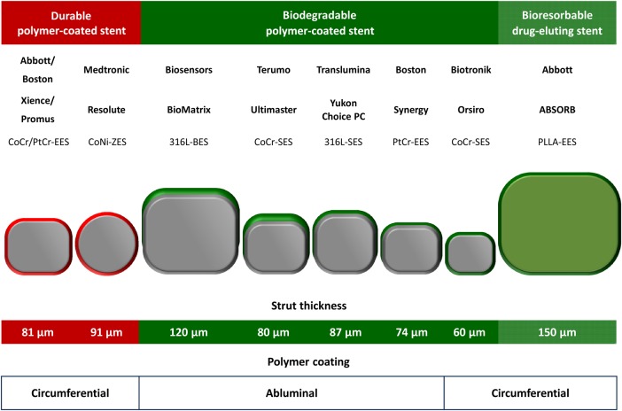
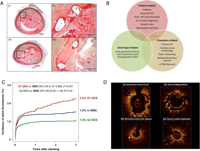
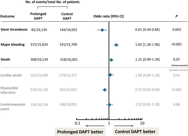
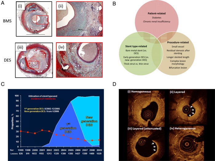
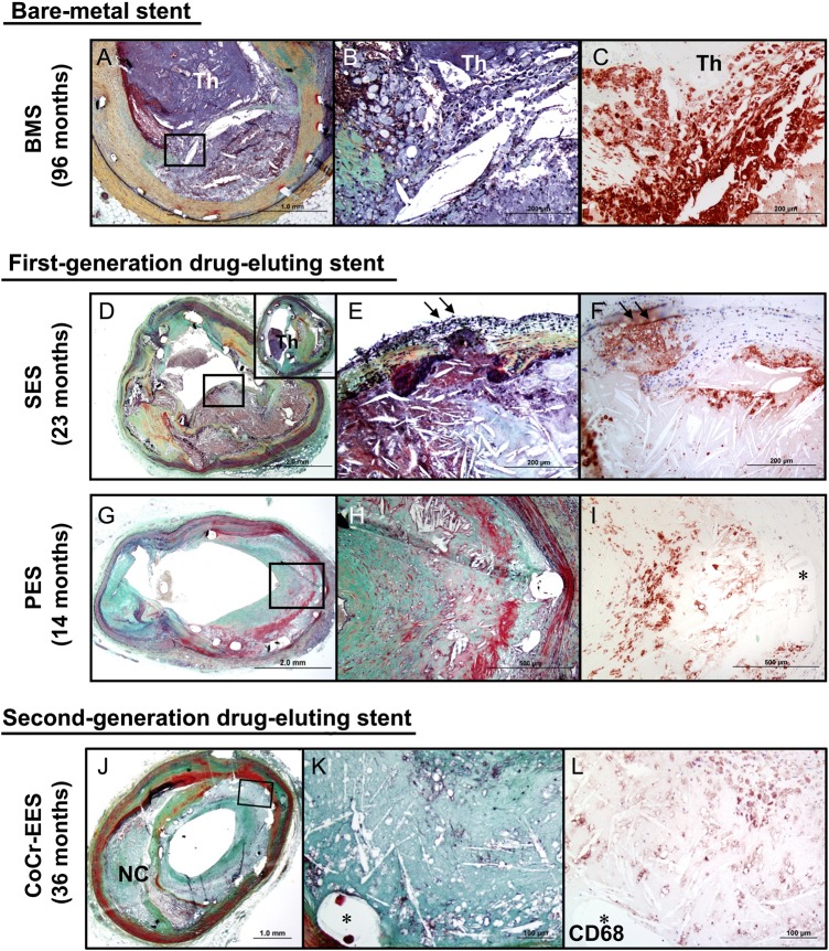
Modern-day stenting procedures leverage advances in pharmacotherapy and device innovation. Patients treated with contemporary antiplatelet agents, peri-procedural antithrombin therapy and new-generation drug-eluting stents (DES) have excellent outcomes over the short to medium term. Indeed, coupled with the reducing costs of these devices in most countries there remain very few indications where patients should be denied treatment with standard-of-care DES therapy. The two major causes of stent failure are stent thrombosis (ST) and in-stent restenosis (ISR). The incidence of both has reduced considerably in recent years. Current clinical registries and randomized trials with broad inclusion criteria show rates of ST at or <1% after 1 year and ∼0.2-0.4% per year thereafter; rates of clinical ISR are 5% respectively. Angiographic surveillance studies in large cohorts show rates of angiographic ISR of ∼10% with new-generation DES. The advent of high-resolution intracoronary imaging has shown that in many cases of late stent failure neoatherosclerotic change within the stented segment represents a final common pathway for both thrombotic and restenotic events. In future, a better understanding of the pathogenesis of this process may translate into improved late outcomes. Moreover, the predominance of non-stent-related disease as a cause of subsequent myocardial infarction during follow-up highlights the importance of lifestyle and pharmacological interventions targeted at modification of the underlying disease process. Finally, although recent developments focus on strategies which circumvent the need for chronically indwelling stents--such as drug-coated balloons or fully bioresorbable stents-more data are needed before the wider use of these therapies can be advocated.

Keywords: Bioresorbable stents; Coronary artery disease; Drug-eluting stents; In-stent restenosis; Neoatherosclerosis; Stent thrombosis.


Figures

Figure 1
Figure 1
Historical perspectives on the development of percutaneous coronary intervention and coronary stenting. The first coronary angioplasty in an awake human was performed by Andreas Grünzig (A) on 16 September 1977 using a balloon catheter fashioned on his kitchen table (B). The patient had a high-grade stenosis of the proximal left anterior descending artery and the initial and late follow-up result (D) was very satisfactory. The first coronary stents implanted in man were performed by Ulrich Sigwart (E) in Lausanne and Jaques Puel (F) in Toulouse in March and April 1986. An angiogram from an initial patient shows high grade stenosis of the proximal left anterior descending artery (G), which was treated with a bare metal Wallstent (H) with a good acute result (I).
Figure 2
Figure 2
Overview of principal characteristics of selected current generation durable polymer and biodegradable polymer drug-eluting stents and fully bioresorbable drug-eluting stents with published large-scale randomized controlled trial data. BES, biolimus-eluting stent; CoCr, cobalt chromium; CoNi, cobalt Nickel; EES, everolimus-eluting stent; PtCr, platinum chromium; SES, sirolimus-eluting stent; ZES, zotarolimus-eluting stent.
Figure 3
Figure 3
Stent thrombosis: central illustration of histopathology, risk factors, incidence, and intravascular imaging features. (A) Representative case showing late stent thrombosis with uncovered struts following drug-eluting stent implantation. Histologic section from a 47-year-old male who had overlapped drug-eluting stents (paclitaxel-eluting stent in the proximal segment and sirolimus-eluting stent in the distal segment) implanted 10 months prior to death. A low-power image (i) shows a platelet-rich occlusive thrombus in the lumen in paclitaxel-eluting stent. A high-power image (ii) of boxed area in (i) shows uncovered struts with peri-strut fibrin. Image (iii) also shows a platelet-rich occlusive thrombus. A high-power image (iv) of boxed area in (iii) shows partially covered struts with neointima (Asterisk indicates stent strut.). (B) Principal risk factors for stent thrombosis classified according to patient-related, stent type-related, and procedure-related risk factors. (C) Incidence of stent thrombosis after bare metal stents, early-generation drug-eluting stents (G1 DES), and new-generation drug-eluting stents (G2 DES); adapted from Tada et al. (D) Representative optical coherence tomography findings from patients presenting with stent thrombosis: (i) persistent uncovered stent struts late after implantation; (ii) marked stent malapposition in the target vessel, this may have been present at the time of implantation or acquired due to late positive remodelling; (iii) neoatherosclerotic plaque formation: diffuse low-signal intensity with higher backscatter in deeper neointimal layers may indicate underlying lipid-rich atherosclerotic tissue; (iv) severe stent underexpansion at site of overlap of multiple stent layers.
Figure 4
Figure 4
Summary results of meta-analysis of trials investigating prolonged duration vs. standard duration dual antiplatelet therapy after drug-eluting stent implantation. Odds ratio with (95% confidence interval) associated with prolonged vs. control dual antiplatelet therapy accounting for events occurred at the longest follow-up available in each included studies. The diamonds and the horizontal lines indicate the odds ratio and the (95% confidence interval) derived from meta-analysis. DAPT, dual antiplatelet therapy; figure based on analysis of data from Cassese et al.
Figure 5
Figure 5
In-stent restenosis: central illustration of histopathology, risk factors, incidence, and intravascular imaging features. (A) Representative histopathological cases showing in-stent restenosis after coronary stenting: (i) low-power magnification of in-stent restenosis in a bare metal stent; (ii) high-power magnification shows predominance of smooth muscle cell-rich neointimal; (iii) low-power magnification of in-stent restenosis in a sirolimus-eluting stent; (iv) higher magnification shows a stent strut with surrounding proteoglycan-rich neointimal tissue and presence of foam cells and neovascularization. (B) Risk factors for stent thrombosis classified according to patient-related, stent type-related, and procedure-related risk factors. (C) Proportion of patients treated with bare metal stents, early-generation drug-eluting stents, and new-generation drug-eluting stents over time and rates of binary angiographic restenosis (red line) in a large registry of patients with angiographic surveillance after stent implantation. Adapted from Cassese et al. (D) Optical coherence tomography imaging of patients with in-stent restenotic tissue during surveillance after stenting; tissue with homogeneous-signal intensity (i) is typical after bare metal stenting; heterogeneous, attenuated, or layered signal intensity tissue (ii–iv) is typical after drug-eluting stents.
Figure 6
Figure 6
Representative cases showing neoatherosclerotic change following bare-metal stent, first-generation drug-eluting stent, and second-generation drug-eluting stent implantation. (AC) Histologic section from a 47-year-old male who had a bare metal stents implanted 8 years prior to death. Note occlusive thrombus in the lumen and ruptured plaque (boxed area in A), which is shown at higher magnification in (B) with large number of macrophages within the lumen as well as at the ruptured cap. Note large number of CD68-positive macrophages at the site of rupture (C). (DF) Histological sections from a 59-year-old male with sirolimus-eluting stents implanted for 23 months who died from stent thrombosis (D). Note thin-cap fibroatheroma with fibrous cap disruption in (E) (arrows) from boxed area in (D). The thrombus was more apparent in the distal section (D; inset). (F) CD68-positive macrophages in the fibrous cap and in the underlying necrotic core. (GI) Histologic sections from a 65-year-old woman with a paclitaxel-eluting stent implanted in the left circumflex artery 14 months antemortem, who died of traumatic brain injury. A low-power image shows a patent lumen with moderate neointimal growth (G), foamy macrophage infiltration and necrotic core formation with cholesterol clefts is seen at high magnification in (H). (i) Same section as (H) showing CD68-positive macrophages in the neointima. (A)–(I) were reproduced with permission from Nakazawa et al. (JL) Histologic sections from a 73-year-old man with cobalt chromium everolimus-eluting stent implanted in the mid left anterior descending for 3 years. A low-power image (J) (Movat) shows moderate luminal narrowing with moderate neointimal growth (69% stenosis) and underlying fibroatheroma. A high-power image (K) of the boxed area in (J) shows necrotic core formation within the neointima where CD68-positive macrophages are identified (L). (J)–(L) were reproduced with permission from Otsuka et al.

References

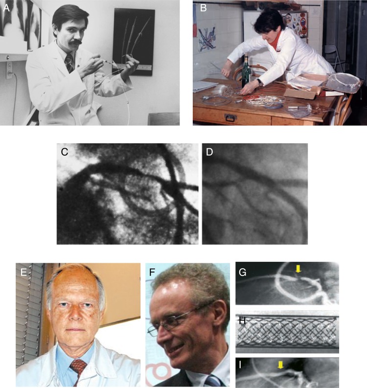
    1. Grüntzig A. Transluminal dilatation of coronary artery stenosis. The Lancet 1978;311:263-263. - PubMed
    1. Meier B. The first patient to undergo coronary angioplasty – 23-year follow-up. N Engl J Med 2001;344:144–145. - PubMed
    1. Monagan D. Journey into the Heart. USA: Gotham Books and Penguin; 2007.
    1. Sigwart U, Puel J, Mirkovitch V, Joffre F, Kappenberger L. Intravascular stents to prevent occlusion and restenosis after transluminal angioplasty. N Engl J Med 1987;316:701–706. - PubMed
    1. Serruys PW, de Jaegere P, Kiemeneij F, Macaya C, Rutsch W, Heyndrickx G, Emanuelsson H, Marco J, Legrand V, Materne P, Belardi J, Sigwart U, Colombo A, Goy JJ, van den Heuvel P, Delcan J, Marie-angele Morel for the Benestent Study Group. A comparison of balloon-expandable-stent implantation with balloon angioplasty in patients with coronary artery disease. Benestent Study Group. N Engl J Med 1994;331:489–495. - PubMed

Publication types

MeSH terms

Substances