. 2015 Oct 13;112(41):12812-7.
doi: 10.1073/pnas.1507094112. Epub 2015 Sep 28.

Endothelial miR-17∼92 cluster negatively regulates arteriogenesis via miRNA-19 repression of WNT signaling

Affiliations

Endothelial miR-17∼92 cluster negatively regulates arteriogenesis via miRNA-19 repression of WNT signaling

Shira Landskroner-Eiger et al. Proc Natl Acad Sci U S A. .

Abstract

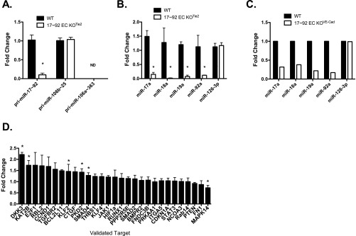
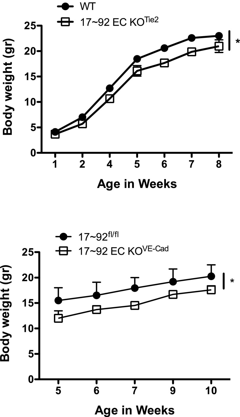
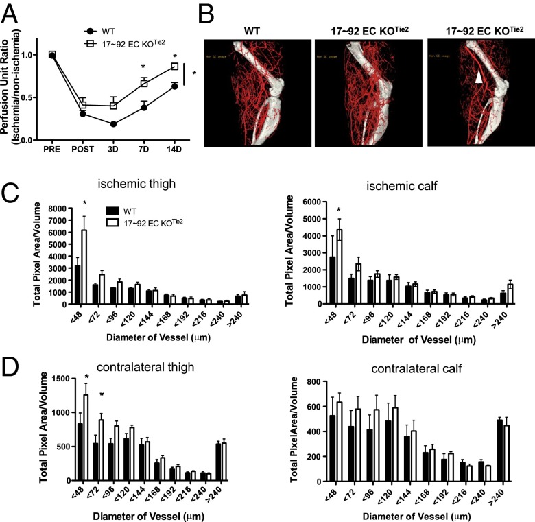
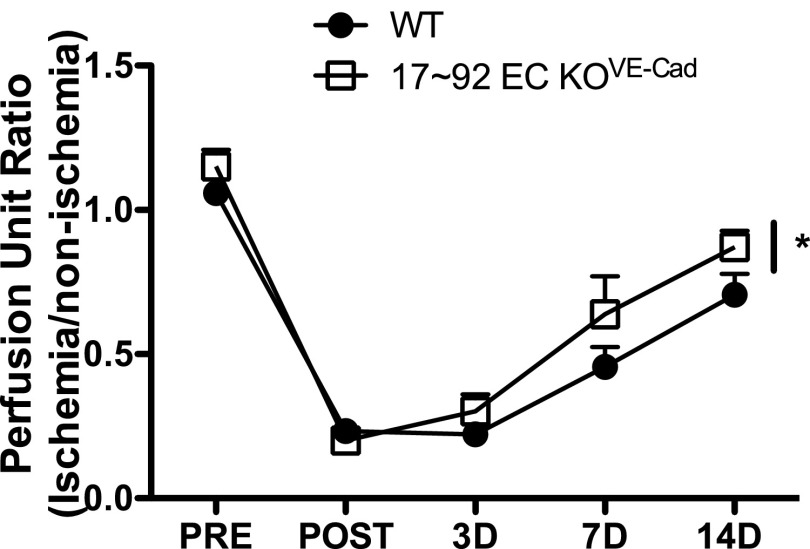
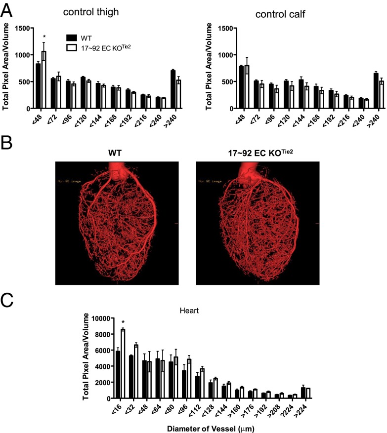
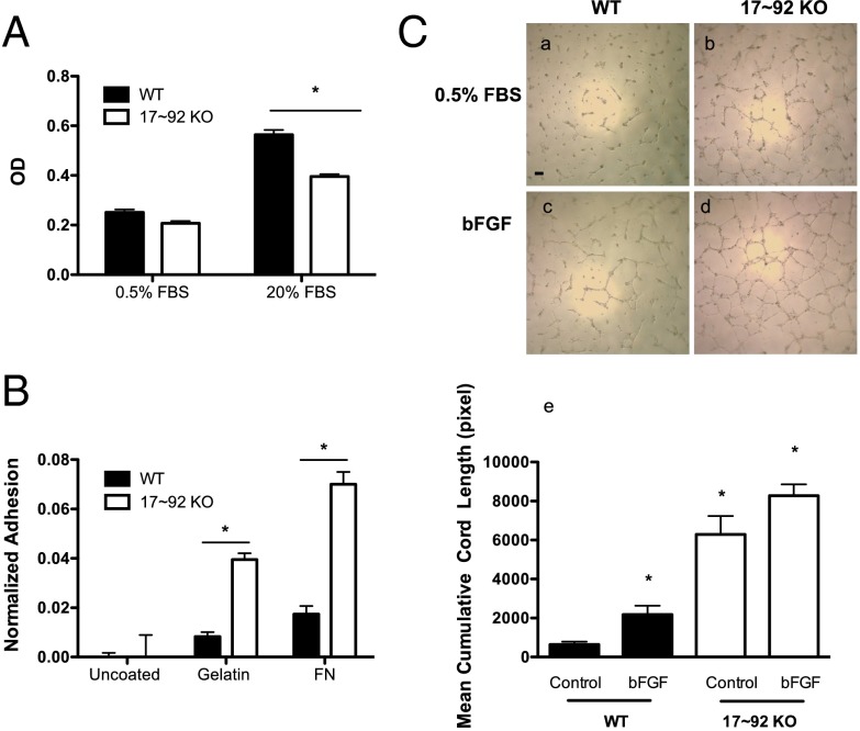
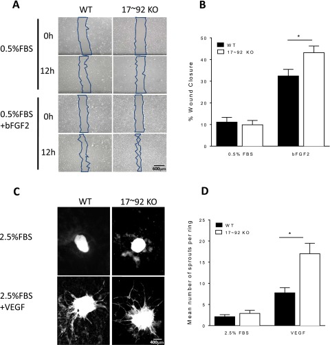
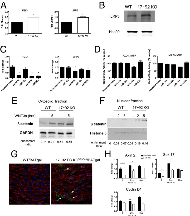
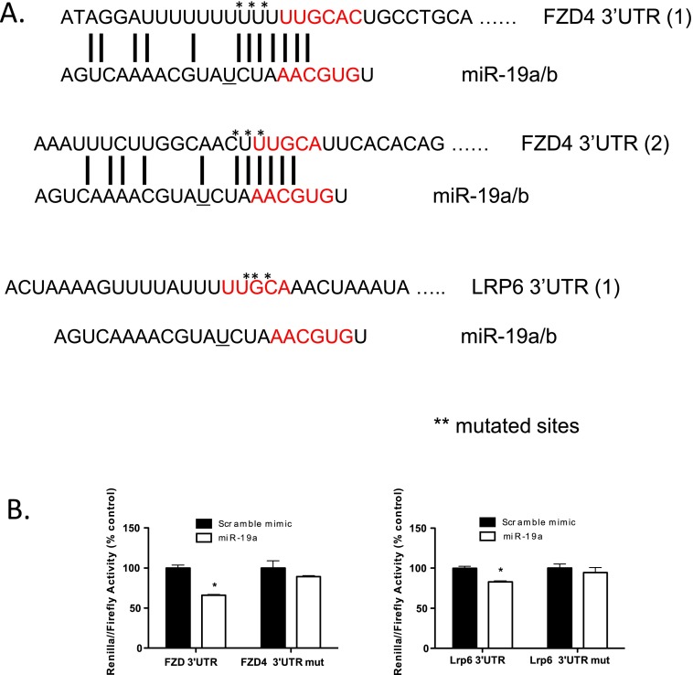
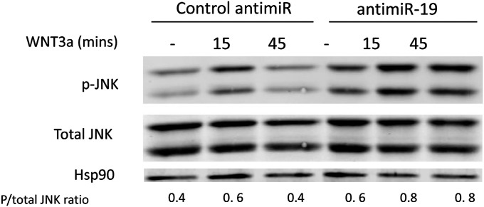
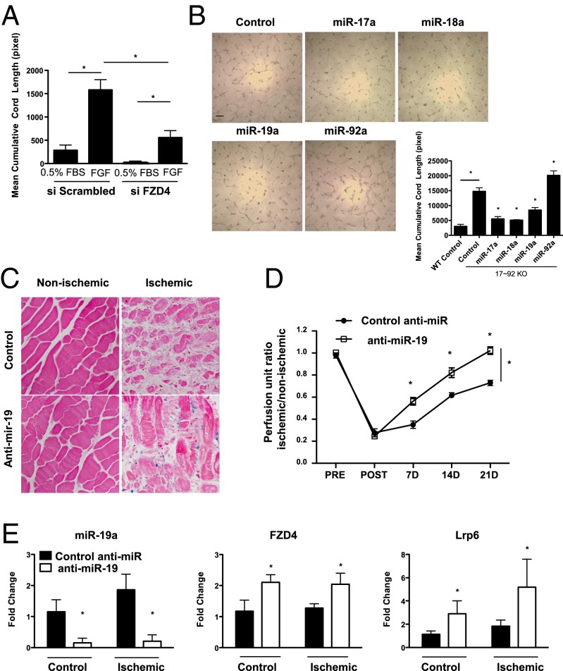
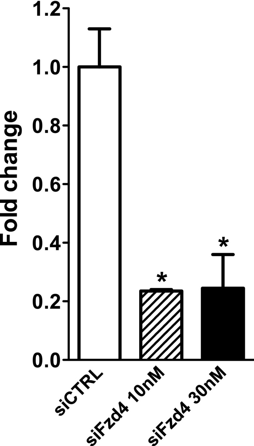
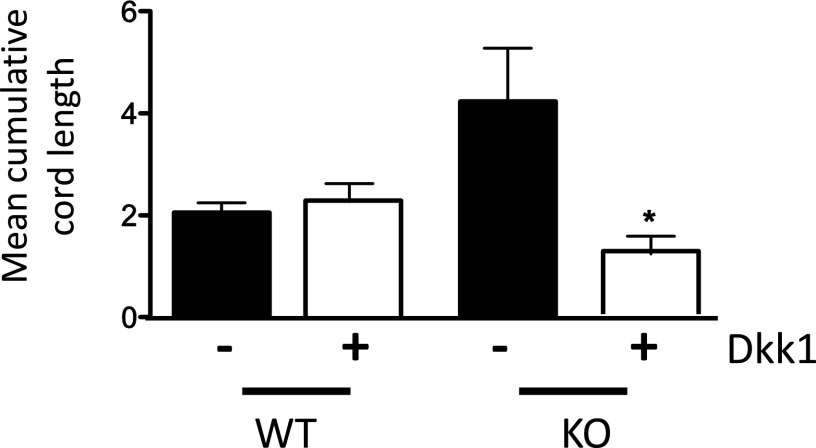
The contribution of endothelial-derived miR-17∼92 to ischemia-induced arteriogenesis has not been investigated in an in vivo model. In the present study, we demonstrate a critical role for the endothelial-derived miR-17∼92 cluster in shaping physiological and ischemia-triggered arteriogenesis. Endothelial-specific deletion of miR-17∼92 results in an increase in collateral density limbs and hearts and in ischemic limbs compared with control mice, and consequently improves blood flow recovery. Individual cluster components positively or negatively regulate endothelial cell (EC) functions in vitro, and, remarkably, ECs lacking the cluster spontaneously form cords in a manner rescued by miR-17a, -18a, and -19a. Using both in vitro and in vivo analyses, we identified FZD4 and LRP6 as targets of miR-19a/b. Both of these targets were up-regulated in 17∼92 KO ECs compared with control ECs, and both were shown to be targeted by miR-19 using luciferase assays. We demonstrate that miR-19a negatively regulates FZD4, its coreceptor LRP6, and WNT signaling, and that antagonism of miR-19a/b in aged mice improves blood flow recovery after ischemia and reduces repression of these targets. Collectively, these data provide insights into miRNA regulation of arterialization and highlight the importance of vascular WNT signaling in maintaining arterial blood flow.

Keywords: arteriogenesis; endothelium; microRNA; vascular.


Conflict of interest statement

The authors declare no conflict of interest.

Figures

Fig. S1.
Fig. S1.
Characterization of miR-17∼92 expression and its validated target gene expression in primary ECs derived from 17∼92 EC KOTie2 and 17∼92 EC KOVE-Cad mice. (A and B) qRT-PCR analysis of cultured primary ECs demonstrating a marked reduction of precursor miR-17∼92 (pri) and its mature derivatives. Data are normalized to WT primary ECs and represent the average of three independent isolations. *P < 0.05. (C) qRT-PCR of primary ECs derived from 17∼92 EC KOVE-Cad animals for detection of mature components of miR-17∼92 and its paralogs. Data are normalized to WT primary ECs and are derived from freshly isolated heart ECs pooled from three animals per group. (D) Expression of validated targets of miR-17∼92 examined via qRT-PCR in primary ECs from WT and 17∼92 EC KOTie2 mice. Data represent fold change compared with primary WT ECs and are derived from at least three independent isolations. *P < 0.05.
Fig. S2.
Fig. S2.
Body weight is reduced in 17∼92 EC KOTie2 and 17∼92 EC KOVE-Cad mice compared with WT mice. Deletion in 17∼92 EC KOVE-Cad was induced at postnatal day 1. n = 4–11 animals per group. *P < 0.05, two-way ANOVA.
Fig. 1.
Fig. 1.
Loss of miR-17∼92 in ECs improves blood flow recovery after HLI. (A) Laser Doppler analysis was used to measure blood flow in the gastrocnemius muscle before, immediately after, and at 3, 7, and 14 d after HLI. n = 9–10 per group. *P < 0.05, two way ANOVA. (B) Reconstructed micro-CT images of arterial vessels of WT and 17∼92 EC KOTie2 mice at 14 d post-HLI. Some 30% of the 17∼92 EC KOTie2 mice had an alternative major collateral artery that was obvious immediately on arteriectomy of the femoral artery, as depicted in C (arrow). (C and D) Quantitative analysis of micro-CT images in the upper and lower limb under ischemic (C) and nonischemic contralateral limbs (D). Data are presented as total number of vascular structures in z-axis slices. n = 4 mice per group. *P < 0.05, two-way ANOVA. All data are mean ± SEM.
Fig. S3.
Fig. S3.
miR-17∼92 deletion in 17∼92 EC KOVE-Cad mice improves HLI blood flow recovery. Deletion was induced at 2 wk before surgery. n = 5–7 per group. *P < 0.05, two-way ANOVA.
Fig. 2.
Fig. 2.
Mice lacking endothelial miR-17∼92 display enhanced arterial density of small arteries in limbs (A: thigh, Left; calf, Right) and heart (B and C). (B) Reconstructed micro-CT images of hearts of WT (Left) and 17∼92 EC KOTie2 (Right) mice show increased arteriole density in heart vessels with a diameter of ≤16 μm. Data are mean ± SEM; n = 4 mice per group. *P < 0.05, two-way ANOVA.
Fig. 3.
Fig. 3.
Characterization of EC phenotypes in ECs from WT and 17∼92 KO mice. (A) BrdU incorporation was used to evaluate proliferation of WT and 17∼92 KO ECs under 0.5% FBS or 20% FBS. *P < 0.05. (B) Adhesion of ECs from both groups was assessed at 30 min after seeding on uncoated, gelatin-coated, or fibronectin-coated plates. *P < 0.05. (C) Cord formation of WT and 17∼92 KO ECs was examined under basal conditions (0.5% FBS) or with bFGF (10 ng/mL) as a stimulant. The 17∼92 KO cells exhibited augmented cord formation under both basal and stimulated conditions. Data are mean ± SEM and are representative of an experiment repeated three additional times and conducted in triplicate. (Scale bar: 200 μm.)
Fig. S4.
Fig. S4.
(A and B) Loss of miR 17∼92 KO in ECs augments recovery after wounding (A) and aortic sprouting (B) in vitro. Primary WT or 17∼92 KO ECs were starved, then wounded and treated with DMEM plus 0.5% FBS or 10 ng/mL bFGF2 for 12 h. The wound area was measured in captured images at 0 and 12 h, and migration was quantified. Data are duplicates from two experiments. (C) Aortic rings from WT and 17∼92VE-Cad mice were embedded in collagen type 1 and treated with +2.5% (vol/vol) FBS or VEGF 100 ng/mL for 6 d. At day 6, rings were fixed with PFA (4%) and stained with BS1 Lectin-FITC to delineate ECs. (D) Images of ring sprouting were obtained and quantified. Data are representative of five or six mice per group, and sprouts from eight or nine rings per mouse were quantified. Error bars represent mean ± SEM. *P < 0.05, one-way ANOVA with Bonferroni’s posttest. Data are representative of an experiment repeated three times and conducted in triplicate. *P < 0.05.
Fig. 4.
Fig. 4.
FZD4 and LRP6 are direct miR-19 targets. (A) qRT-PCR analysis showing enhanced mRNA expression of FZD4 and LRP6 in 17∼92 KO ECs compared with WT ECs. Data are from four independent isolations. (B) Expression levels of LRP6 protein in ECs isolated from 17∼92 KO and WT mice. (C) Changes in mRNA expression levels of FZD4 and Lrp6 in 17∼92 KO ECs following transfection with the indicated mimics (30 nM). (D) Cotransfection of miR-19a (60 nM) significantly reduced 3′ UTR luciferase reporter activity for FZD4 and LRP6 measured in HEK293 cells. The data shown are representative of an experiment repeated three times and conducted in triplicate. (E and F) Primary ECs were treated with either control CM (labeled –) or WNT3a CM for 5 h or for 2 or 5 h. Fractionated cytoplasmic (E) and nuclear (F) samples were analyzed for protein stabilization of β-catenin. Data are quantified below the blots relative to loading controls and are representative of an experiment repeated three times. (G) BAT-gal reporter for WNT/β-catenin signaling was examined in WT/BAT-gal or 17∼92 EC KOVE-Cad/BAT animals. Adult mice were killed at 2 wk after tamoxifen induction of cluster deletion, and mesenteric arteries were triple-stained en face for β-gal (green) to detect β-catenin–induced expression, for PECAM (red) and DAPI. Double-staining for β-gal and PECAM was detected in the nuclei of 17∼92 KO animals; n = 3–4. (Scale bar: 25 μm.) (H) mRNA expression of WNT3a transcriptionally regulated genes as analyzed by qRT-PCR. ECs were transfected with scramble or mimic miR-19a (30 nM). At 48 h after transfection, cells were serum-starved for 3 h and then stimulated with control CM or WNT3a CM for 2–6 h. Data are presented as fold change compared with WT ECs, mean ± SEM. *P < 0.05.
Fig. S5.
Fig. S5.
LRP6 and FZD4 are functionally relevant miR-19 target genes. (A) Schematic representation of the sequences of the miR-19 predicted binding sites in the 3′ UTR of FZD4 and LRP6. Mutation sites are marked with an asterisk. Sequence differences between miR19a and miR19b are indicated by “_”. (B) Mutation of predicted miR-19a target sites reduces miR-19a– mediated repression of 3′ UTR LUC activity of FZD4 and LRP6. Data are mean ± SEM from three independent experiments.
Fig. S6.
Fig. S6.
Anti–miR-19 augments noncanonical WNT stimulation of JNK. MLECs were transfected with control or anti–miR-19 (60 nM each) for 48 h before Wnt3a stimulation. Cells were starved for 4 h, then treated with Wnt3a-conditioned medium for the indicated times. Lysates were collected, run on SDS/PAGE gel, and immunoblotted for p-JNK, total JNK, and Hsp90.
Fig. 5.
Fig. 5.
FZD4 knockdown and miR-17∼92 cluster components rescue augmented cord formation in cluster-deficient ECs, and LNA–miR-19 improves flow recovery. (A) The 17∼92 KO ECs transfected with siFZD4 (20 nM) or control siRNA were plated on Matrigel to assess cord formation. *P < 0.05. (B) Individual components of the cluster were transfected into 17∼92 KO ECs, and cord formation was quantified. *P < 0.05. n = 3 experiments in triplicate. (Scale bar: 200 μm.) (C and D) Aged BAT-gal or WT mice were injected with 12.5 mg/kg LNA–anti–miR-19 or LNA scramble control for 3 consecutive days before induction of HLI, followed by injections on days 1, 8, and 15 post-HLI. (C) β-catenin–dependent gene expression (LacZ) in ischemic limbs. (D) Blood flow changes; n = 9 mice per group. *P < 0.05, two way ANOVA. (E) qRT-PCR analysis of thigh muscle tissue confirming miR-19 repression in animals treated with the LNA-modified anti–miR-19. Sample analyses depict overall significant increases in FZD4 (Middle) and LRP6 (Right) mRNA levels in mice treated with LNA miR-19 expression compared with control LNA-miR–treated mice. n = 4 mice per group. *P < 0.05, two-way ANOVA. All data are mean ± SEM.
Fig. S7.
Fig. S7.
Efficient deletion of FZD4 in 17∼92 KO ECs. The 17∼92 KO ECs were transfected with 10 nM or 30 nM siFZD4. After 48 h, cells were harvested, and mRNA levels of FZD4 were measured by qRT-PCR. The data are representative of an experiment repeated three times and conducted in triplicate. *P < 0.05.
Fig. S8.
Fig. S8.
DKK1 antagonizes cord formation in ECs from miR-17–92 KO mice. WT and miR 17∼92 KO ECs were placed into cord assays in the presence of 0.5% serum and untreated or treated with Dkk1 (1 mg/mL). Cord formation was monitored over 5–8 h. Data are representative of an experiment conducted in triplicate and subsequently repeated, showing that DKK1 reduces cord formation in KO ECs in a dose-dependent manner. Data are presented as mean ± SEM. *P < 0.05.

References

    1. Bartel DP. MicroRNAs: Target recognition and regulatory functions. Cell. 2009;136(2):215–233. - PMC - PubMed
    1. Xiao C, et al. Lymphoproliferative disease and autoimmunity in mice with increased miR-17-92 expression in lymphocytes. Nat Immunol. 2008;9(4):405–414. - PMC - PubMed
    1. Suárez Y, et al. Dicer-dependent endothelial microRNAs are necessary for postnatal angiogenesis. Proc Natl Acad Sci USA. 2008;105(37):14082–14087. - PMC - PubMed
    1. Dews M, et al. Augmentation of tumor angiogenesis by a Myc-activated microRNA cluster. Nat Genet. 2006;38(9):1060–1065. - PMC - PubMed
    1. Ventura A, et al. Targeted deletion reveals essential and overlapping functions of the miR-17 through 92 family of miRNA clusters. Cell. 2008;132(5):875–886. - PMC - PubMed

Publication types

MeSH terms