

Clinicomycological Study of Dermatophytosis in South India
- PMID: 26417157
- PMCID: PMC4559634
- DOI: 10.4103/0974-2727.163135
Clinicomycological Study of Dermatophytosis in South India
Abstract
Introduction: Dermatophytic infections are commonly encountered a problem and constitute more than 50% of cases in dermatology outpatient departments. Diagnosis of these infections requires the proper use of laboratory methods.
Objectives: This study was conducted to know the etiology of dermatophytosis in patients attending Tertiary Care Level Hospital in South India and to compare the efficacy of Sabouraud's dextrose agar (SDA) with actidione and dermatophyte test medium (DTM) in isolating and identifying dermatophytes.
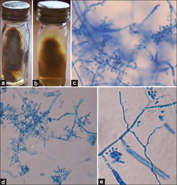
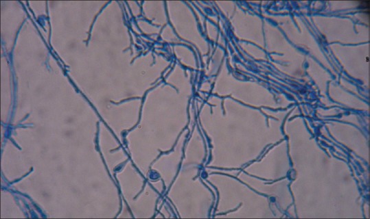
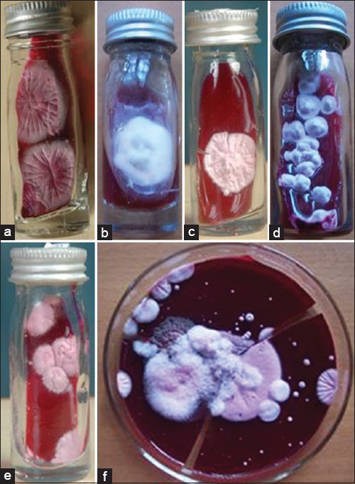
Materials and methods: A total of 110 samples which included 101 skin samples and 9 hair samples from clinically suspected dermatophytosis were collected. Direct microscopy by KOH and culture on SDA with actidione and DTM were done.
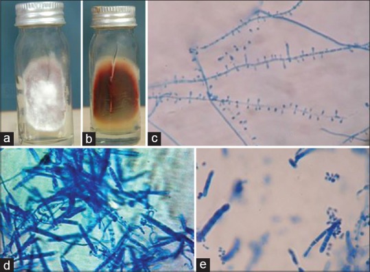
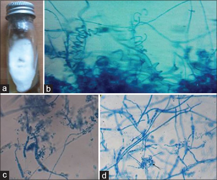
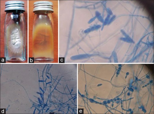
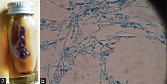
Results: Of 110 samples collected, 58.18% were KOH positive for fungal filaments and 56.36% were culture positive for dermatophytes. More number of cases were observed between age groups of 21-40 years. Males were more affected compared to females. Tinea corporis was the common clinical presentation observed (40%). Trichophyton rubrum (58.06%) was the predominant isolate recovered in all clinical presentations but Trichophyton violaceum was the most common isolate in tinea capitis. All culture positives were grown on both SDA with actidione and DTM. Appearance of growth was earlier on DTM that is, within 10 days compared to SDA with actidione where growth started appearing only after 10 days. This is statistically significant P < 0.0001 (χ(2) = 71.6). Species level identification on primary isolation was possible when grown on SDA with actidione and it was not possible with the growth on DTM on primary isolation.
Conclusion: DTM is a good screening medium in laboratory diagnosis of dermatophytosis when compared to SDA with actidione. But DTM is inferior to SDA with actidione in identification of dermatophyte species.
Keywords: Dermatophte test medium; Sabouraud's dextrose agar with actidione; dermatophytosis.
Figures
References
-
- Koneman EW, Allen SD, Janda WM, Schreckenberber P, Win WC., Jr . 8th ed. Philadelphia: JB Lippincott; 1997. Color Atlas and Textbook of Diagnostic Microbiology.
-
- Mohanty JC, Mohanty SK, Sahoo RC, Sahoo A, Praharaj N. Incidence of dermatophytosis in Orissa. Indian J Med Microbiol. 1998;16:78–80.
-
- Sentamilselvi G, Kamalam A, Ajithadas K, Janaki C, Thambiah AS. Scenario of chronic dermatophytosis: An Indian study. Mycopathologia. 1997;140:129–35. - PubMed
-
- Singh S, Beena PM. Profile of dermatophyte infections in Baroda. Indian J Dermatol Venereol Leprol. 2003;69:281–3. - PubMed
LinkOut - more resources
Full Text Sources
Other Literature Sources