. 2015 Aug 13;6(9):3352-61.
doi: 10.1364/BOE.6.003352. eCollection 2015 Sep 1.

Periscope for noninvasive two-photon imaging of murine retina in vivo

Affiliations

Periscope for noninvasive two-photon imaging of murine retina in vivo

Patrycjusz Stremplewski et al. Biomed Opt Express. .

Abstract

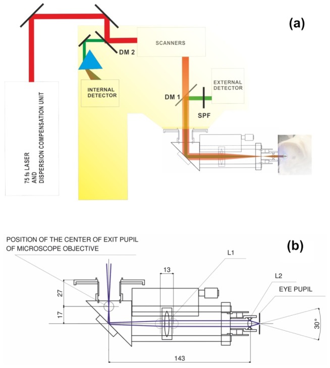
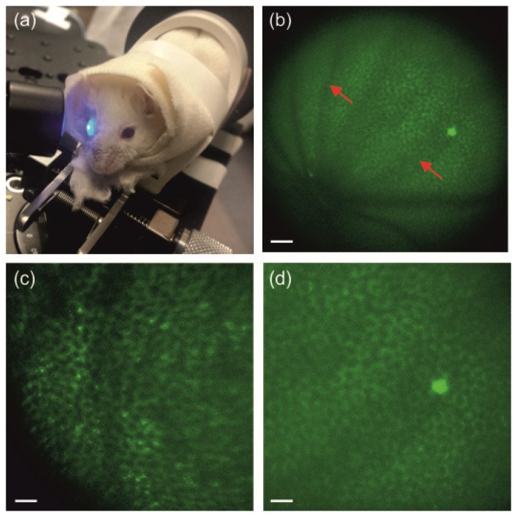
Two-photon microscopy allows visualization of subcellular structures in the living animal retina. In previously reported experiments it was necessary to apply a contact lens to each subject. Extending this technology to larger animals would require fitting a custom contact lens to each animal and cumbersome placement of the living animal head on microscope stage. Here we demonstrate a new device, periscope, for coupling light energy into mouse eye and capturing emitted fluorescence. Using this periscope we obtained images of the RPE and their subcellular organelles, retinosomes, with larger field of view than previously reported. This periscope provides an interface with a commercial microscope, does not require contact lens and its design could be modified to image retina in larger animals.

Keywords: (170.0110) Imaging systems; (170.4460) Ophthalmic optics and devices; (170.5755) Retina scanning; (180.4315) Nonlinear microscopy; (330.7327) Visual optics, ophthalmic instrumentation.


Figures

Fig. 1
Fig. 1
(a) Position of the periscope in the microscope setup. DM 1 and DM 2 are dichroic mirrors, SPF is a short pass filter. (b) Schematic of the periscope and centers of the extreme rays (blue lines). L1 is an achromatic doublet with a 75 mm focal length and 25 mm diameter; L2 is an achromatic doublet with a 10 mm focal length and an 8 mm diameter. Unless otherwise specified, all dimensions shown are in millimeters.
Fig. 2
Fig. 2
(a) Calculated imaging depth adjustment range. The central position of L1 results in a collimated periscope output beam and a focal spot located 2.5 mm from eye lens. (b) Calculated FOV versus position of focal spot with respect to the cardinal plane of the eye lens.
Fig. 3
Fig. 3
Two-photon imaging of the RPE in living WT and Rpe65–/– mice. (a) Mouse eye illuminated with the newly designed periscope. (b) Image of the RPE in the WT albino mouse. (c), (d). Enlarged images of the RPE in Rpe65–/– (c) and WT (d) albino mice. Excitation wavelength: 730 nm. Mean power at the cornea: 20 mW in (c) and 25 mW in (b) and (d). Scale bars: 100 µm in (b), and 50 µm in (c) and (d). Red arrows indicate shadows from retinal vasculature.
Fig. 4
Fig. 4
Images of retinal capillaries in a live WT mouse after tail vain injection with FITC-BSA. Images were acquired with a TPM equipped with a periscope. Top row images were acquired with dispersion compensation (DC), bottom row images were procured without dispersion compensation. Excitation wavelengths are indicated in each image. Scale bars are equal to 100 µm.
Fig. 5
Fig. 5
Fluorescence emission spectrum from the RPE of a live Rpe65−/−mouse. (a) Spectrum measured with a periscope had maximum at 524 nm. (b) Deconvolution of the spectrum (black squares, red line) revealed two components (green lines), one with a maximum at 524 nm and the second, a long wavelength shoulder with a maximum at 627 nm.

References

    1. Wang B. G., Koenig K., Riemann I., Krieg R., Halbhuber K. J., “Intraocular multiphoton microscopy with subcellular spatial resolution by infrared femtosecond lasers,” Histochem. Cell Biol. 126(4), 507–515 (2006). 10.1007/s00418-006-0187-0 - DOI - PubMed
    1. Han M., Giese G., Schmitz-Valckenberg S., Bindewald-Wittich A., Holz F. G., Yu J., Bille J. F., Niemz M. H., “Age-related structural abnormalities in the human retina-choroid complex revealed by two-photon excited autofluorescence imaging,” J. Biomed. Opt. 12(2), 024012 (2007). 10.1117/1.2717522 - DOI - PubMed
    1. Bueno J. M., Gualda E. J., Artal P., “Adaptive optics multiphoton microscopy to study ex vivo ocular tissues,” J. Biomed. Opt. 15(6), 066004 (2010). 10.1117/1.3505018 - DOI - PubMed
    1. Gualda E. J., Bueno J. M., Artal P., “Wavefront optimized nonlinear microscopy of ex vivo human retinas,” J. Biomed. Opt. 15(2), 026007 (2010). 10.1117/1.3369001 - DOI - PubMed
    1. Bueno J. M., Giakoumaki A., Gualda E. J., Schaeffel F., Artal P., “Analysis of the chicken retina with an adaptive optics multiphoton microscope,” Biomed. Opt. Express 2(6), 1637–1648 (2011). 10.1364/BOE.2.001637 - DOI - PMC - PubMed