. 2015 Aug 21;6(9):3462-74.
doi: 10.1364/BOE.6.003462. eCollection 2015 Sep 1.

In vivo microscopy of hemozoin: towards a needle free diagnostic for malaria

Affiliations

In vivo microscopy of hemozoin: towards a needle free diagnostic for malaria

Jennifer L Burnett et al. Biomed Opt Express. .

Abstract

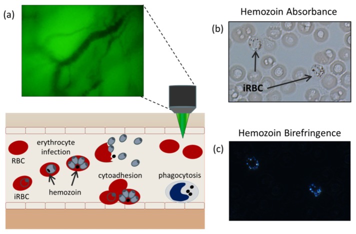
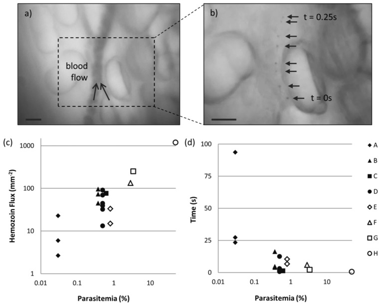
Clinical diagnosis of malaria suffers from poor specificity leading to overtreatment with antimalarial medications. Alternatives, like blood smear microscopy or antigen-based tests, require a blood sample. We investigate in vivo microscopy as a needle-free malaria diagnostic. Two optical signatures, birefringence and absorbance, of the endogenous malaria by-product hemozoin were evaluated as in vivo optical biomarkers. Hemozoin birefringence was difficult to detect in highly scattering tissue; however, hemozoin absorbance was observed in increasingly complex biological environments and detectable over a clinically-relevant range of parasitemia in vivo in a P. yoelii-infected mouse model of malaria.

Keywords: (170.0180) Microscopy; (170.1470) Blood or tissue constituent monitoring; (260.1440) Birefringence.


Figures

Fig. 1
Fig. 1
(a) Diagnostic concept: malaria-infected cells are detected circulating through the superficial microvasculature using in vivo microscopy. (b) The malaria parasite by-product, hemozoin, visualized under bright-field and (c) cross-polarized illumination.
Fig. 2
Fig. 2
(a) Schematic diagram of the MVM with three illumination modes: TR, transmission red, TG, transmission green, and XP, cross polarized reflectance. (b) USAF 1951 resolution target imaged under TR and XP (inset). (c) The measured and calculated magnification of the MVM as a function of working distance.
Fig. 3
Fig. 3
MVM imaging of multilayer phantom. (a) Negative control blood sample imaged under TR and (b) XP. (c) P. yoelii-infected blood sample imaged under TR and (d) XP. Arrows indicate hemozoin particles. Scale bars = 20 µm. (e) Temporal variations in SBR quantified for 10 colocalized ROIs. (f) The SBR as a function of increasing epithelial scattering by increasing Intralipid concentration.
Fig. 4
Fig. 4
Ex vivo imaging of microvasculature in excised mouse tissue from (a-c) a negative control animal and (d-i) a P. yoelii-infected animal. Arrows indicate hemozoin particles. Scale bars = 20 µm.
Fig. 5
Fig. 5
In vivo imaging of circulating blood in superficial microvasculature of mouse model. (a,b) Axial translation of focal plane in TG mode in P. yoelii-infected animal. (c,d) Selected frames from TR mode video (Visualization 1) collected at imaging site outlined in (b). (e) Imaging of non-infected animal in TG mode and (f) TR mode. Scale bars = 20 µm.
Fig. 6
Fig. 6
In vivo imaging of circulating hemozoin over a range of parasitemia in mouse model. (a) Vessel located using TG mode. (b) Overlaid frames from correlating TR mode Visualization 2 show single hemozoin tracked over 0.25 s. Scale bars = 20 µm. (c) Hemozoin flux plotted as a function of parasitemia according to subject (A-H). (d) Estimation of time to detect one hemozoin structure as a function of parasitemia.

References

    1. World Health Organization, World Malaria Report (WHO Press, 2012).
    1. Rafael M. E., Taylor T., Magill A., Lim Y. W., Girosi F., Allan R., “Reducing the burden of childhood malaria in Africa: the role of improved,” Nature 444(Suppl 1), 39–48 (2006).10.1038/nature05445 - DOI - PubMed
    1. Rowe J. A., Claessens A., Corrigan R. A., Arman M., “Adhesion of Plasmodium falciparum-infected erythrocytes to human cells: molecular mechanisms and therapeutic implications,” Expert Rev. Mol. Med. 11, e16 (2009).10.1017/S1462399409001082 - DOI - PMC - PubMed
    1. Egan T. J., “Recent advances in understanding the mechanism of hemozoin (malaria pigment) formation,” J. Inorg. Biochem. 102(5-6), 1288–1299 (2008).10.1016/j.jinorgbio.2007.12.004 - DOI - PubMed
    1. Wilson B. K., Behrend M. R., Horning M. P., Hegg M. C., “Detection of malarial byproduct hemozoin utilizing its unique scattering properties,” Opt. Express 19(13), 12190–12196 (2011).10.1364/OE.19.012190 - DOI - PubMed

LinkOut - more resources