. 2015 Nov;42(10):2833-42.
doi: 10.1111/ejn.13088. Epub 2015 Nov 2.

The anterior paraventricular thalamus modulates neuronal excitability in the suprachiasmatic nuclei of the rat

Affiliations

The anterior paraventricular thalamus modulates neuronal excitability in the suprachiasmatic nuclei of the rat

Javier Alamilla et al. Eur J Neurosci. 2015 Nov.

Abstract

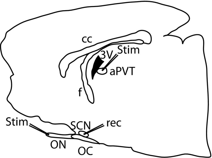
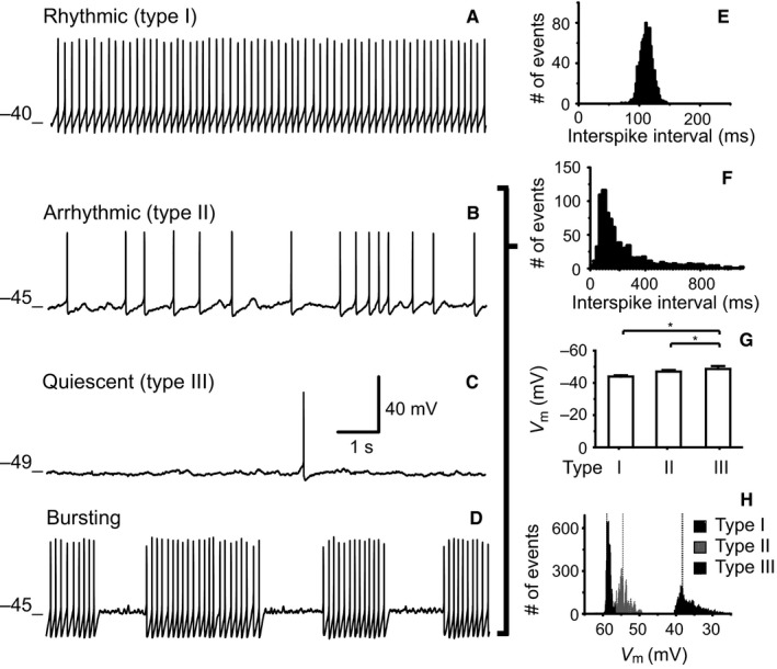
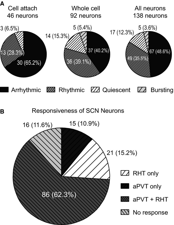
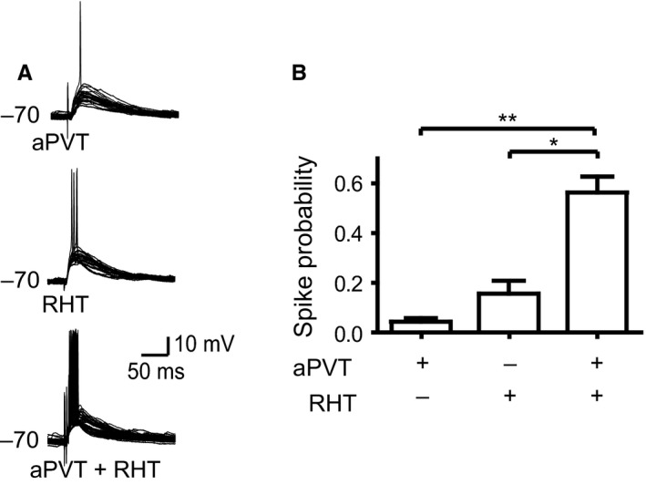
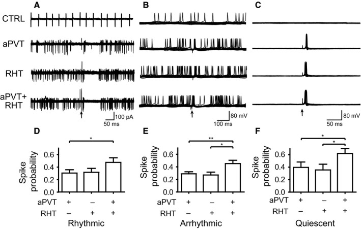
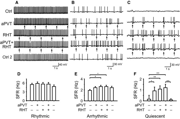
The suprachiasmatic nucleus (SCN) in mammals is the master clock which regulates circadian rhythms. Neural activity of SCN neurons is synchronized to external light through the retinohypothalamic tract (RHT). The paraventricular thalamic nucleus (PVT) is a neural structure that receives synaptic inputs from, and projects back to, the SCN. Lesioning the anterior PVT (aPVT) modifies the behavioral phase response curve induced by short pulses of bright light. In order to study the influence of the aPVT on SCN neural activity, we addressed whether the stimulation of the aPVT can modulate the electrical response of the SCN to either retinal or RHT stimulation. Using in vitro and in vivo recordings, we found a large population of SCN neurons responsive to the stimulation of either aPVT or RHT pathways. Furthermore, we found that simultaneous stimulation of the aPVT and the RHT increased neuronal responsiveness and spontaneous firing rate (SFR) in neurons with a low basal SFR (which also have more negative membrane potentials), such as quiescent and arrhythmic neurons, but no change was observed in neurons with rhythmic firing patterns and more depolarized membrane potentials. These results suggest that inputs from the aPVT could shift the membrane potential of an SCN neuron to values closer to its firing threshold and thus contribute to integration of the response of the circadian clock to light.

Keywords: circadian; hypothalamus; intralaminar nuclei; light entrainment; patch-clamp.


Figures

Figure 1
Figure 1
Schematic representation of the preparation. A sagittal slice used for patch‐clamp recordings. The stimulation electrodes (stim) were located on the aPVT and the ON, whereas the recording electrode (rec) was placed into the SCN. Abbreviations: cc, corpus callosum; f, fornix, 3V, third ventricle; aPVT, anterior paraventricular thalamus; ON, optic nerve; OC, optic chiasm; SCN, suprachiasmatic nucelus.
Figure 2
Figure 2
Location of extracellular recording marks in vivo. (A) Neurons recorded outside the SCN, circles on the right indicate the neurons that did not respond to stimulation; on the left, are neurons that responded to only retinal stimulation, to only aPVT stimulation and to both stimuli. (B) Neurons recorded in the SCN. Abbreviations: OC, optic chiasm; AHy, anterior hypothalamic area; RCh, retrochiasmatic area; mpo, medial preoptic area; LPO, lateral preoptic area; 3V, third ventricle; AH, anterior hypothalamus; F, fornix; PVN, paraventricular nucleus of the hypothalamus; LH, lateral hypothalamus.
Figure 3
Figure 3
Examples of 3 SCN neurons recorded in vivo on which functional convergence of the retina and the aPVT occurs. (A) Peristimulus histogram showing a SCN neuron responding to aPVT but not to retinal stimulation; when both afferents were stimulated, the response to aPVT stimulation was modulated from an excitation to an inhibition. (B) CUSUM analysis (cumulative frequency analysis) from the same neurons as in A. (C and D) Two additional examples of the CUSUM analysis; in C, the neuron responded to aPVT stimulation with an excitation that disappears when the aPVT is stimulated simultaneously with the retina. In D, the SCN neuron responded to retinal stimulation with an excitation followed by inhibition; simultaneous stimulation of both the aPVT and the retina modulates the response to inhibition‐excitation‐inhibition. The rectangles in the CUSUM graphs indicate the statistically significant responses (Mann‐Whitney test, P < 0.05).
Figure 4
Figure 4
Schematic representation of 122 SCN neurons recorded in vitro which responded to different types of stimulation; the inset indicates the outline of sagittal section shown in Fig. 1. Abbreviations: OC, optic chiasm; mpo, medial preoptic area; LA, lateral anterior hypothalamus.
Figure 5
Figure 5
Firing patterns recorded in SCN neurons in vitro. (A) Rhythmic firing presents a narrow Gaussian interspike interval distribution (E). (B) Arrhythmic firing in which the interspike interval has a Poisson distribution (F). (C) Quiescent, with sporadic action potentials. (D) Bursting firing in which bursts of action potentials are followed by periods of silence. Voltage traces A–C indicate the in Vm for the different spike patterns shown. (E) Interspike interval histogram of the rhythmic neuron shown in A. The histogram has a normal distribution, characteristic of this type of spike pattern. (F) Interspike interval histogram of the arrhythmic neuron illustrated in B. This type of distribution is also characteristic of the arrhythmic, the quiescent and the bursting spike patterns. (G) Average and SEM of the Vm related to the different spike patterns of all SCN neurons recorded in whole‐cell mode, *P < 0.05. (H) Frequency histograms of raw data (20 000 points) belonging to rhythmic neurons (type I), arrhythmic neurons (type II) and quiescent neurons (type III). Dotted lines show the median for each distribution.
Figure 6
Figure 6
(A) Relative frequency of spike patterns found in the SCN in cell‐attached (left), whole‐cell (middle) and all patch‐clamp (right) recordings. (B) Relative distribution of responsiveness of SCN neurons to aPVT and RHT stimulation, as well as the non‐responsive neurons recorded in vitro (cell‐attached and whole‐cell modes).
Figure 7
Figure 7
SS of the aPVT and the RHT increases the probability of firing action potentials in hyperpolarized SCN neurons. (A) Overlaid voltage traces (30 sweeps) displaying occurrence of the action potentials by aPVT, RHT and concurrent aPVT and RHT stimulation (SS). (B) SS induces a significant increase in the firing probability over individual stimulation (*P < 0.05; **P < 0.001).
Figure 8
Figure 8
SS of the RHT and the aPVT increases the probability of firing action potentials in different neuronal firing patterns. Overlaid traces (10 sweeps) illustrate different spike patterns and action potential induction by individual afferent stimulation and concurrent stimulation in (A) rhythmic neurons (cell‐attached recordings), (B) arrhythmic neurons and (C) quiescent neurons. The arrows indicate the stimulation of the afferents. (D–F) Modulation of firing probability in (D) rhythmic neurons (*P < 0.05), (E) arrhythmic neurons (*P < 0.01; **P < 0.05) and (F) quiescent neurons (*P < 0.05).
Figure 9
Figure 9
SS has different effects on firing frequency depending on the spontaneous firing pattern and SFR. The afferent stimulation is indicated on the left. Firing patterns and changes induced in firing rate during different stimulations are shown for (A and D) rhythmic neurons, (B and E) arrhythmic neurons (*P < 0.05) and (C and F) quiescent neurons (*P < 0.05; **P < 0.01; ***P < 0.001). Arrows indicate the moment of afferent stimulation.

References

    1. Alamilla, J. & Aguilar‐Roblero, R. (2010) Glutamate and GABA neurotransmission from the paraventricular thalamus to the suprachiasmatic nuclei in the rat. J. Biol. Rhythm, 25, 28–36. - PubMed
    1. Alamilla, J. , Perez‐Burgos, A. , Quinto, D. & Aguilar‐Roblero, R. (2014) Circadian modulation of the Cl(‐); Equilibrium potential in the rat suprachiasmatic nuclei. BioMed Res. Int., 2014, 15. - PMC - PubMed
    1. Belle, M.D. , Diekman, C.O. , Forger, D.B. & Piggins, H.D. (2009) Daily electrical silencing in the mammalian circadian clock. Science, 326, 281–284. - PubMed
    1. Bos, N.P. & Mirmiran, M. (1993) Effects of excitatory and inhibitory amino acids on neuronal discharges in the cultured suprachiasmatic nucleus. Brain Res. Bull., 31, 67–72. - PubMed
    1. Cavalcante, J.S. , Costa, M.S. , Santee, U.R. & Britto, L.R. (2005) Retinal projections to the midline and intralaminar thalamic nuclei in the common marmoset (Callithrix jacchus). Brain Res., 1043, 42–47. - PubMed

Publication types

LinkOut - more resources