

Structural and Functional Characterization of the Hazelnut Allergen Cor a 8
- PMID: 26417906
- PMCID: PMC4616228
- DOI: 10.1021/acs.jafc.5b03534
Structural and Functional Characterization of the Hazelnut Allergen Cor a 8
Abstract
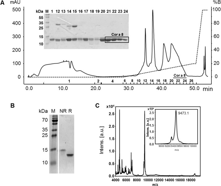
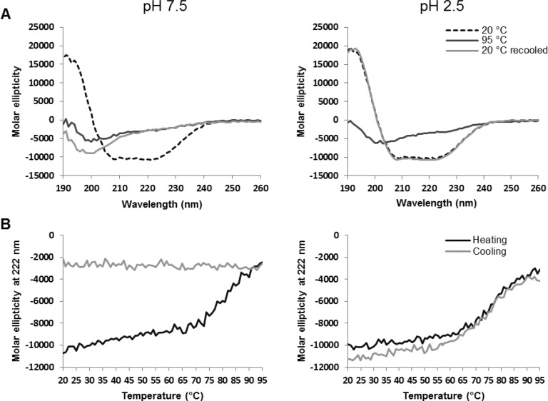
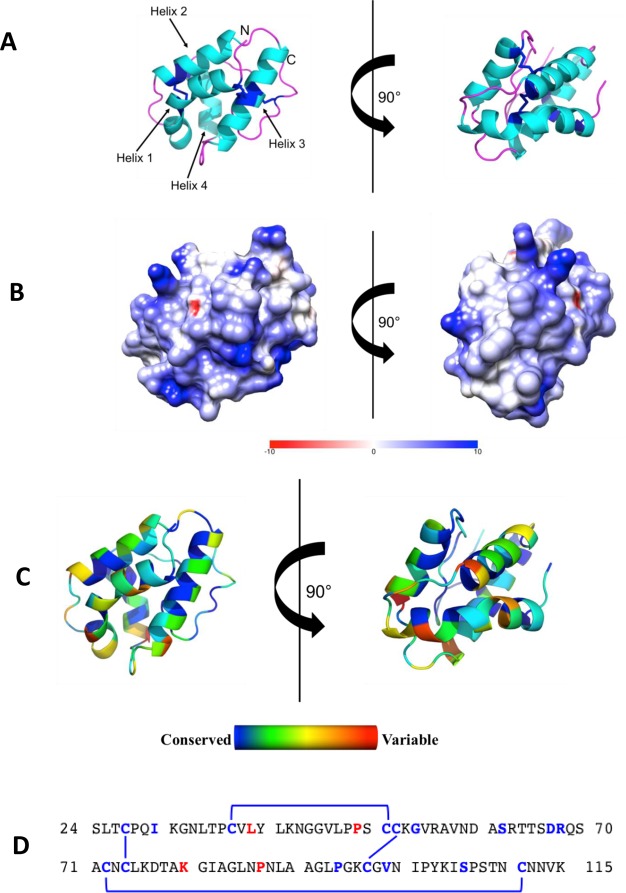
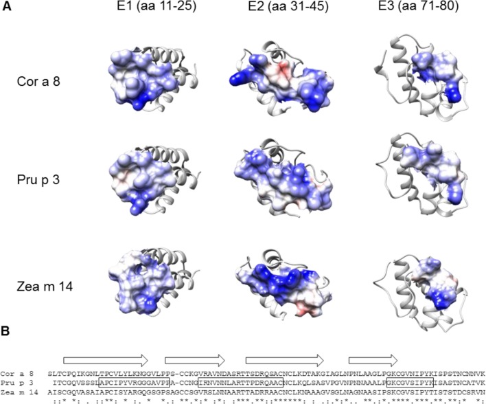
Nonspecific lipid transfer proteins (nsLTPs) are basic proteins, stabilized by four disulfide bonds, and are expressed throughout the plant kingdom. These proteins are also known as important allergens in fruits and tree nuts. In this study, the nsLTP from hazelnuts, Cor a 8, was purified and its crystal structure determined. The protein is stable at low pH and refolds after thermal denaturation. Molecular dynamics simulations were used to provide an insight into conformational changes of Cor a 8 upon ligand binding. When known epitope areas from Pru p 3 were compared to those of Cor a 8, differences were obvious, which may contribute to limited cross-reactivity between peach and hazelnut allergens. Differences in epitope regions may contribute to limited cross-reactivity between Cor a 8 and nsLTPs from other plant sources. The structure of Cor a 8 represents the first resolved structure of a hazelnut allergen.
Keywords: allergen; food allergy; hazelnut; nonspecific lipid transfer protein; protein crystallization.
Conflict of interest statement
The authors declare no competing financial interest.
Figures
References
-
- Muraro A.; Werfel T.; Hoffmann-Sommergruber K.; Roberts G.; Beyer K.; Bindslev-Jensen C.; Cardona V.; Dubois A.; duToit G.; Eigenmann P.; Fernandez Rivas M.; Halken S.; Hickstein L.; Host A.; Knol E.; Lack G.; Marchisotto M. J.; Niggemann B.; Nwaru B. I.; Papadopoulos N. G.; Poulsen L. K.; Santos A. F.; Skypala I.; Schoepfer A.; Van Ree R.; Venter C.; Worm M.; Vlieg-Boerstra B.; Panesar S.; de Silva D.; Soares-Weiser K.; Sheikh A.; Ballmer-Weber B. K.; Nilsson C.; de Jong N. W.; Akdis C. A. EAACI food allergy and anaphylaxis guidelines: diagnosis and management of food allergy. Allergy 2014, 69, 1008–1025. 10.1111/all.12429. - DOI - PubMed
-
- De Knop K. J.; Verweij M. M.; Grimmelikhuijsen M.; Philipse E.; Hagendorens M. M.; Bridts C. H.; De Clerck L. S.; Stevens W. J.; Ebo D. G. Age-related sensitization profiles for hazelnut (Corylus avellana) in a birch-endemic region. Ped. Allergy Immunol. 2011, 22, e139–149. 10.1111/j.1399-3038.2011.01112.x. - DOI - PubMed
-
- Hansen K. S.; Ballmer-Weber B. K.; Sastre J.; Lidholm J.; Andersson K.; Oberhofer H.; Lluch-Bernal M.; Ostling J.; Mattsson L.; Schocker F.; Vieths S.; Poulsen L. K. Component-resolved in vitro diagnosis of hazelnut allergy in Europe. J. Allergy Clin. Immunol. 2009, 123, 1134–1141. 10.1016/j.jaci.2009.02.005. - DOI - PubMed
Publication types
MeSH terms
Substances
Associated data
- Actions
LinkOut - more resources
Full Text Sources
Other Literature Sources
Molecular Biology Databases
