. 2015 Oct 6;6(30):28988-98.
doi: 10.18632/oncotarget.4902.

Changes in the gene expression of co-cultured human fibroblast cells and osteosarcoma cells: the role of microenvironment

Affiliations

Changes in the gene expression of co-cultured human fibroblast cells and osteosarcoma cells: the role of microenvironment

Viviana Salvatore et al. Oncotarget. .

Abstract

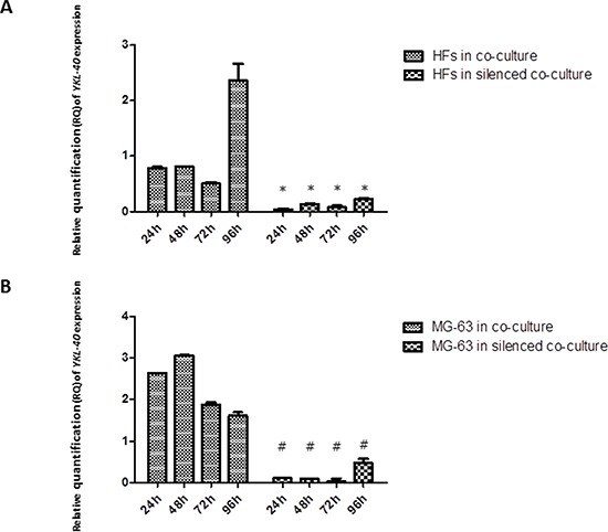
Background: The progression of malignant tumors does not depend exclusively on the autonomous properties of cancer cells; it is also influenced by tumor stroma reactivity and is under strict microenvironmental control. By themselves, stromal cells are not malignant, and they maintain normal tissue structure and function. However, through intercellular interactions or by paracrine secretions from cancer cells, normal stromal cells acquire abnormal phenotypes that sustain cancer cell growth and tumor progression. In their dysfunctional state, fibroblast and immune cells produce chemokines and growth factors that stimulate cancer cell growth and invasion. In our previous work, we established an in vitro model based on a monolayer co-culture system of healthy human fibroblasts (HFs) and human osteosarcoma cells (the MG-63 cell line) that simulates the microenvironment of tumor cells and healthy cells. The coexistence between MG-63 cells and HFs allowed us to identify the YKL-40 protein as the main marker for verifying the influence of tumor cells grown in contact with healthy cells.

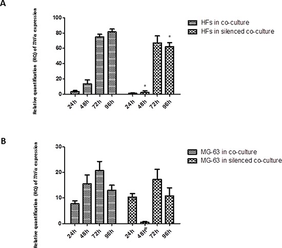
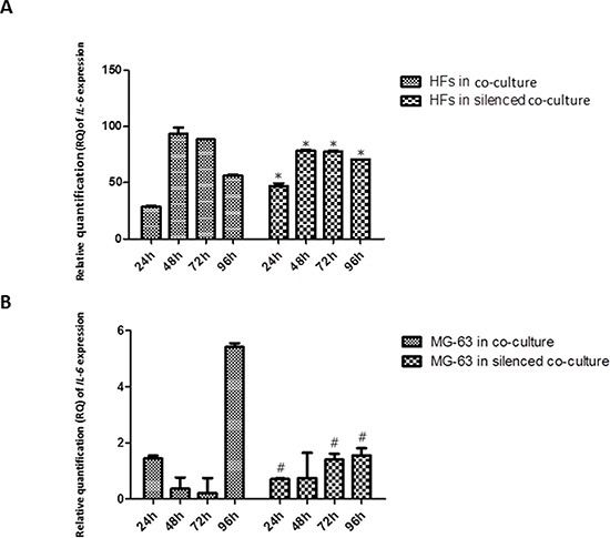
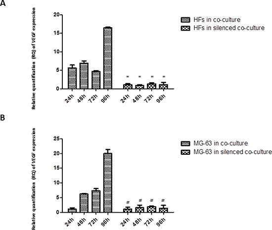
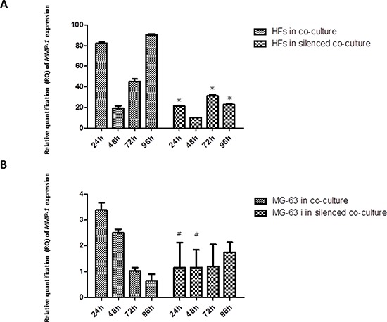
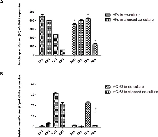
Methods: In this study, we evaluated the interactions of HFs and MG-63 cells in a transwell co-culture system over 24 h, 48 h, 72 h, and 96 h. We analyzed the contributions of these populations to the tumor microenvironment during cancer progression, as measured by multiple markers. We examined the effect of siRNA knockdown of YKL-40 by tracking the subsequent changes in gene expression within the co-culture. We validated the expression of several genes, focusing on those involved in cancer cell invasion, inflammatory responses, and angiogenesis: TNF alpha, IL-6, MMP-1, MMP-9, and VEGF. We compared the results to those from a transwell co-culture without the YKL-40 knockdown.

Results: In a pro-inflammatory environment promoted by TNF alpha and IL-6, siRNA knockdown of YKL-40 caused a down-regulation of VEGF and MMP-1 expression in HFs.

Conclusions: These findings demonstrated that the tumor microenvironment has an influence on the gene expression of healthy surrounding tissues and on the process of tumorigenicity and it is emerging as attractive targets for therapeutic strategies.

Keywords: YKL-40; inflammation; osteosarcoma cells; siRNA; tumor microenvironment.


Conflict of interest statement

CONFLICTS OF INTEREST

The authors declare no financial interest or sources of research funding which could affect integrity of the scientific work presented.

Figures

Figure 1
Figure 1. A. and B
Real-Time PCR for TNF-α. The gene expression levels of TNF-α had a similar trend in co-culture and co-culture with silenced YKL-40: TNF-α mRNA increased significantly in HFs, reaching the highest level at 72 h and 96 h, whereas in the tumor cells, it remained constant and low. * represents a significant difference from HF cells grown in normal co-culture, P < 0.05. # represents a significant difference from MG-63 cells grown in normal co-culture, P < 0.05.
Figure 2
Figure 2. A. and B
Real-Time PCR for IL-6. IL-6 mRNA had high expression in HFs from co-culture and co-culture with silenced YKL-40. MG-63 cells in both co-culture conditions poorly expressed IL-6. * represents a significant difference from HF cells grown in normal co-culture, P < 0.05. # represents a significant difference from MG-63 cells grown in normal co-culture, P < 0.05.
Figure 3
Figure 3. A. and B
Real-Time PCR for YKL-40. In MG-63 cells, siRNA knockdown of YKL-40 reached the maximum efficiency at 72 h, with respect to the expression of YKL-40 in normally co-cultured MG-63 cells, in which the levels of YKL-40 are significantly higher. In normal co-culture, fibroblasts had increased levels of YKL-40, reaching the highest peak at 96 h. In fibroblasts co-cultured with the silenced MG-63 cells, YKL-40 was at the minimum level. * represents a significant difference from HF cells grown in normal co-culture, P < 0.05. # represents a significant difference from MG-63 cells grown in normal co-culture, P < 0.05.
Figure 4
Figure 4. A. and B
Real-Time PCR for VEGF. VEGF expression in fibroblasts and MG-63 cells in normal co-culture continued to increase through 96 h. In the YKL-40-silenced co-culture, the levels of VEGF were low in both cell populations. * represents a significant difference from HF cells grown in normal co-culture, P < 0.05. # represents a significant difference from MG-63 cells grown in normal co-culture, P < 0.05.
Figure 5
Figure 5. A. and B
Real-Time PCR for MMP-1. The expression of MMP-1 was weak in MG-63 cells grown in both co-cultures. In normal co-culture, MMP-1 was strongly expressed in HFs and increased to a maximum peak at 96 h. In the silenced co-culture, its expression was three-fold lower than in the normal co-culture. * represents a significant difference from HF cells grown in normal co-culture, P < 0.05. # represents a significant difference from MG-63 cells grown in normal co-culture, P < 0.05.
Figure 6
Figure 6. A. and B
Real-Time PCR for MMP-9. The expression of MMP-9 tended to decrease steadily as fibroblasts were grown in normal co-culture, whereas in fibroblasts grown in silenced co-culture, it remained fairly constant for at least 72 h, before declining to lower levels by the 96 h time point. The MG-63 cells of both conditions expressed MMP-9 only weakly. * represents a significant difference from HF cells grown in normal co-culture, P < 0.05. # represents a significant difference from MG-63 cells grown in normal co-culture, P < 0.05.

References

    1. Kidd S, Spaeth E, Watson K, Burks J, Lu H, Klopp A, Andreeff M, Marini FC. Origins of the tumor microenvironment: quantitative assessment of adipose-derived and bone marrow-derived stroma. PLoS One. 2012 doi: 10.1371/journal.pone.0030563. - DOI - PMC - PubMed
    1. C Rodrigues-Lisoni F, Peitl P, Vidotto A, Polachini GM, Maniglia JV, Carmona-Raphe J, Cunha BR, Henrique T, Souza CF, Teixeira RAP, Fukuyama EE, Michaluart P, De Carvalho MB, Oliani SM. Genomics and proteomics approaches to the study of cancer-stroma interactions 2, Head and Neck Genome Project BMC Medical Genomics. 2010 doi: 10.1186/1755-8794-3-14. - DOI - PMC - PubMed
    1. Kuperwasser C. The tumor stromal microenvironment as modulator of malignant behavior. J Mammary Gland Biol Neoplasia. 2010 Dec; doi: 10.1007/s10911-010-9198-5. - DOI - PubMed
    1. Balkwill F. TNF-alpha in promotion and progression of cancer. Cancer Metastasis Rev. 2006;25:409–16. - PubMed
    1. Whiteside T. The tumor microenvironment and its role in promoting tumor growth. Oncogene. 2008;27:5904–5912. - PMC - PubMed

Publication types

MeSH terms