

Imaging of carbonic anhydrase IX with an 111In-labeled dual-motif inhibitor
- PMID: 26418876
- PMCID: PMC4741798
- DOI: 10.18632/oncotarget.5254
Imaging of carbonic anhydrase IX with an 111In-labeled dual-motif inhibitor
Abstract
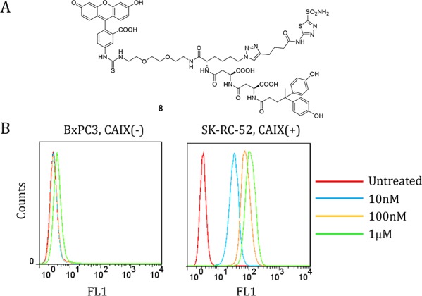
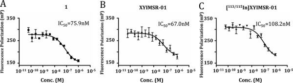
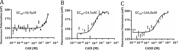
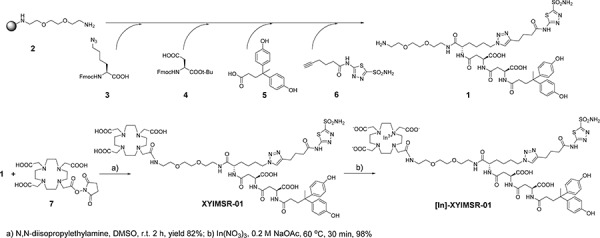
We developed a new scaffold for radionuclide-based imaging and therapy of clear cell renal cell carcinoma (ccRCC) targeting carbonic anhydrase IX (CAIX). Compound XYIMSR-01, a DOTA-conjugated, bivalent, low-molecular-weight ligand, has two moieties that target two separate sites on CAIX, imparting high affinity. We synthesized [111In]XYIMSR-01 in 73.8-75.8% (n = 3) yield with specific radioactivities ranging from 118 - 1,021 GBq/μmol (3,200-27,600 Ci/mmol). Single photon emission computed tomography of [111In]XYIMSR-01 in immunocompromised mice bearing CAIX-expressing SK-RC-52 tumors revealed radiotracer uptake in tumor as early as 1 h post-injection. Biodistribution studies demonstrated 26% injected dose per gram of radioactivity within tumor at 1 h. Tumor-to-blood, muscle and kidney ratios were 178.1 ± 145.4, 68.4 ± 29.0 and 1.7 ± 1.2, respectively, at 24 h post-injection. Retention of radioactivity was exclusively observed in tumors by 48 h, the latest time point evaluated. The dual targeting strategy to engage CAIX enabled specific detection of ccRCC in this xenograft model, with pharmacokinetics surpassing those of previously described radionuclide-based probes against CAIX.
Keywords: CAIX; indium-111; molecular imaging; renal cell carcinoma; single photon emission computed tomography.
Conflict of interest statement
The authors declare no relevant conflicts of interest.
Figures


References
-
- Srigley JR, Delahunt B, Eble JN, Egevad L, Epstein JI, Grignon D, Hes O, Moch H, Montironi R, Tickoo SK, Zhou M, Argani P, Panel IRT. The International Society of Urological Pathology (ISUP) Vancouver Classification of Renal Neoplasia. The American journal of surgical pathology. 2013;37:1469–1489. - PubMed
-
- Siegel RL, Miller KD, Jemal A. Cancer statistics, 2015. CA: a cancer journal for clinicians. 2015;65:5–29. - PubMed
-
- Pichler M, Hutterer GC, Chromecki TF, Jesche J, Kampel-Kettner K, Eberhard K, Hoefler G, Pummer K, Zigeuner R. Trends of stage, grade, histology and tumour necrosis in renal cell carcinoma in a European centre surgical series from 1984 to 2010. Journal of clinical pathology. 2012;65:721–724. - PubMed
-
- Lipworth L, Morgans AK, Edwards TL, Barocas DA, Chang SS, Herrell SD, Penson DF, Resnick MJ, Smith JA, Clark PE. Renal cell cancer histologic subtype distribution differs by race and sex. BJU international. 2014 - PubMed
-
- Umbreit EC, Shimko MS, Childs MA, Lohse CM, Cheville JC, Leibovich BC, Blute ML, Thompson RH. Metastatic potential of a renal mass according to original tumour size at presentation. BJU international. 2012;109:190–194. discussion 194. - PubMed
Publication types
MeSH terms
Substances
Grants and funding
LinkOut - more resources
Full Text Sources
Other Literature Sources
