. 2015 Oct 20;6(32):33878-92.
doi: 10.18632/oncotarget.5302.

BPTF promotes tumor growth and predicts poor prognosis in lung adenocarcinomas

Affiliations

BPTF promotes tumor growth and predicts poor prognosis in lung adenocarcinomas

Meng Dai et al. Oncotarget. .

Abstract

BPTF, a subunit of NURF, is well known to be involved in the development of eukaryotic cell, but little is known about its roles in cancers, especially in non-small-cell lung cancer (NSCLC). Here we showed that BPTF was specifically overexpressed in NSCLC cell lines and lung adenocarcinoma tissues. Knockdown of BPTF by siRNA significantly inhibited cell proliferation, induced cell apoptosis and arrested cell cycle progress from G1 to S phase. We also found that BPTF knockdown downregulated the expression of the phosphorylated Erk1/2, PI3K and Akt proteins and induced the cleavage of caspase-8, caspase-7 and PARP proteins, thereby inhibiting the MAPK and PI3K/AKT signaling and activating apoptotic pathway. BPTF knockdown by siRNA also upregulated the cell cycle inhibitors such as p21 and p18 but inhibited the expression of cyclin D, phospho-Rb and phospho-cdc2 in lung cancer cells. Moreover, BPTF knockdown by its specific shRNA inhibited lung cancer growth in vivo in the xenografts of A549 cells accompanied by the suppression of VEGF, p-Erk and p-Akt expression. Immunohistochemical assay for tumor tissue microarrays of lung tumor tissues showed that BPTF overexpression predicted a poor prognosis in the patients with lung adenocarcinomas. Therefore, our data indicate that BPTF plays an essential role in cell growth and survival by targeting multiply signaling pathways in human lung cancers.

Keywords: BPTF; lung cancer; prognosis; tumor growth.


Conflict of interest statement

CONFLICTS OF INTEREST

The authors declare no conflict of interest.

Figures

Figure 1
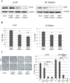
Figure 1. BPTF was overexpressed in NSCLC cell lines and lung adenocarcinoma tissues
A. The expression of BPTF in human normal lung cell HLF and various NSCLC cell lines was analyzed by Western blot. B. The expression of BPTF in human normal lung cell HLF and various NSCLC cell lines was analyzed by RT-PCR. C. The expression of BPTF in human lung adenocarcinoma and their adjacent non-malignant lung tissues from 5 different patients with lung cancers (Case 1, 2, 3, 4 and 5) was detected by immunohistochemical staining (magnification, 200x and 400x).
Figure 2
Figure 2. Knockdown of BPTF inhibited proliferation of NSCLC cells
A. The expression of BPTF in A549 and NCI-H460 cell lines were analyzed by Western blot after transfected with BPTF siRNA. Mock, non treatment; control siRNA, non specific siRNA; BPTF siRNA-1 and BPTF siRNA-2, BPTF specific siRNA. B. Cell viability of A549 and NCI-H460 cells was measured by MTT assay. The mean and SD value got from 3 independent experiments are marked (*P < 0.05; **P < 0.01). C. Colonies (>50 μm) were counted 10–12 days in A549 and NCI-H460 cells after transfected by siRNA. Each bar represents the mean colony number and SD of 3 wells (*P < 0.05; **P < 0.01).
Figure 3
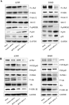
Figure 3. Knockdown of BPTF suppressed MAPK and PI3K-AKT signaling pathways
A. The key protein in MAPK pathways were detected by immunoblot in A549 and NCI-H460 cells 3 days after transfected by siRNA. B. The proteins in PI3K-AKT pathway in A549 and NCI-H460 were analyzed by Western blot 3 days after siRNA transfection.
Figure 4
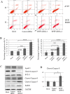
Figure 4. Knockdown of BPTF activated apoptosis by caspase-dependent pathway
A. A549 and NCI-H460 cells transfected with siRNA for 3 days were analyzed by FACS using an Annexin V-FITC/PI-staining kit. B. Apoptosis was calculated in terms of the FITC-positive in cells. Each bar represents the mean and SD value of 3 experiments (**P < 0.01; ***P < 0.001). C. NCI-H460 cells with knockdown of BPTF were analyzed by Western blot with antibodies of Apaf-1, cleaved caspase-9, BCL-2, cleaved caspase-8, caspase-7 and PARP1. D. The quantitative analysis for the cleaved caspase-8.
Figure 5
Figure 5. Knockdown of BPTF inhibited cell cycle
A. BPTF was analyzed by Western blot in A549 and H322 cell lines transfected with BPTF siRNA for 3 days. B. Cell cycle of A549 and H322 cells with knockdown of BPTF was tested by FACS. C. Cell cycle was analyzed in terms of peaks of G1, S and G2/M. Each bar represents the SD from 3 independent experiments. D. Key proteins associated with cell cycle were detected by Western blot.
Figure 6
Figure 6. BPTF predicted a poor prognosis in lung adenocarcinoma
A. Typical examples for negative, weak, moderate and strong BPTF expression in lung adenocarcinoma tissues (Magnification 200X). B. Overall survival of lung adenocarcinoma patients with high or low BPTF expression was analyzed by Kaplan-Meier analysis.
Figure 7
Figure 7. Knockdown of BPTF by shRNA inhibited lung cancer cell growth in vitro and in vivo
A. The expression of BPTF in A549 and NCI-H460 cell lines were analyzed by Western blot after transfected with non-specific control shRNA or BPTF shRNA. B. Cell viability of A549 and NCI-H460 cells was measured by MTT assay. The mean and SD value got from 3 independent experiments are marked (*P < 0.05; **P < 0.01). C. Tumor growth curves in xenografts of A549 cells for each group (n = 5). Dots represent the mean, while bars indicate the SD. (*P < 0.05). D. Tumor graft from the 2 different groups treated with BPTF shRNA and Control shRNA respectively. E. The expression of BPTF, VEGF, P-Erk, P-Akt, P21, BCL-2 and GAPDH were examined from 3 pairs of tumor xenografts in non-specific control shRNA and BPTF-specific shRNA-treated nude mice by Western blot. “C” means control shRNA, and “B”means “BPTF shRNA”.

References

    1. Torre LA, Bray F, Siegel RL, Ferlay J, Lortet-Tieulent J, Jemal A. Global cancer statistics. CA: a cancer journal for clinicians. 2012:2015. - PubMed
    1. Grose D, Devereux G, Milroy R. Comorbidity in lung cancer: important but neglected. a review of the current literature. Clinical lung cancer. 2011;12:207–211. - PubMed
    1. Ryan RJ, Bernstein BE. Molecular biology. Genetic events that shape the cancer epigenome. Science. 2012;336:1513–1514. - PubMed
    1. Neely KE, Workman JL. The complexity of chromatin remodeling and its links to cancer. Biochimica et biophysica acta. 2002;1603:19–29. - PubMed
    1. Bruce Alberts AJ, Julian Lewis, Martin Raff, Keith Roberts, Peter Walter. Molecular Biology of the Cell. 4. New York: Garland Science; 2002.

Publication types

MeSH terms