. 2016 Jan:179:107-15.
doi: 10.1016/j.cbpc.2015.09.009. Epub 2015 Sep 28.

Purification and characterization of tenerplasminin-1, a serine peptidase inhibitor with antiplasmin activity from the coral snake (Micrurus tener tener) venom

Affiliations

Purification and characterization of tenerplasminin-1, a serine peptidase inhibitor with antiplasmin activity from the coral snake (Micrurus tener tener) venom

Jeilyn Vivas et al. Comp Biochem Physiol C Toxicol Pharmacol. 2016 Jan.

Abstract

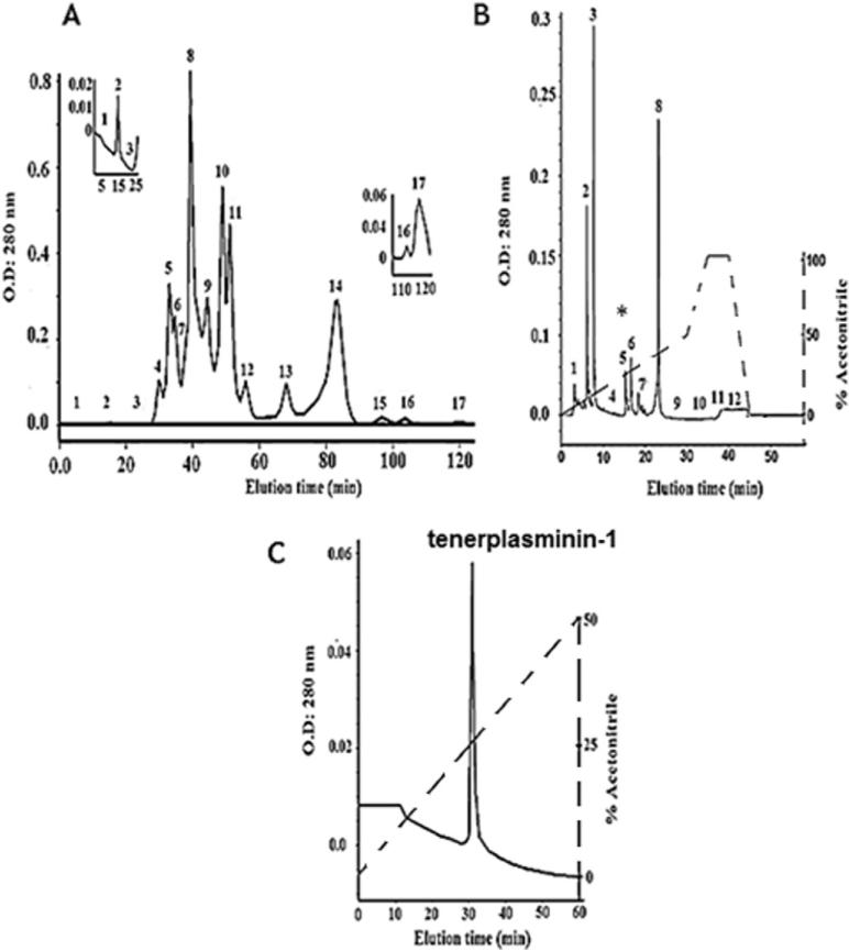
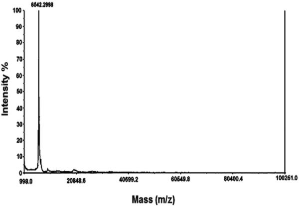
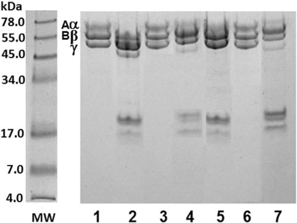
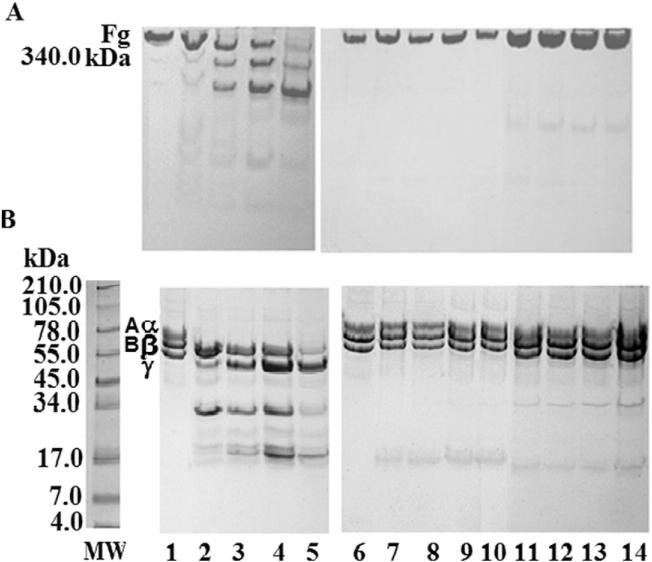
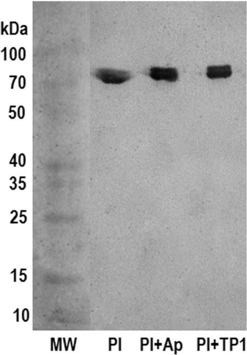
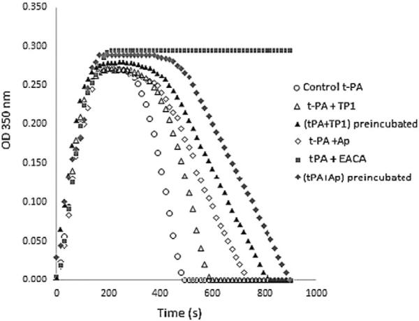
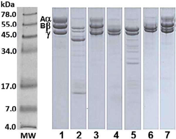
A plasmin inhibitor, named tenerplasminin-1 (TP1), was isolated from Micrurus tener tener (Mtt) venom. It showed a molecular mass of 6542Da, similarly to Kunitz-type serine peptidase inhibitors. The amidolytic activity of plasmin (0.5nM) on synthetic substrate S-2251 was inhibited by 91% following the incubation with TP1 (1nM). Aprotinin (2nM) used as the positive control of inhibition, reduced the plasmin amidolytic activity by 71%. Plasmin fibrinolytic activity (0.05nM) was inhibited by 67% following incubation with TP1 (0.1nM). The degradation of fibrinogen chains induced by plasmin, trypsin or elastase was inhibited by TP1 at a 1:2, 1:4 and 1:20 enzyme:inhibitor ratio, respectively. On the other hand, the proteolytic activity of crude Mtt venom on fibrinogen chains, previously attributed to metallopeptidases, was not abolished by TP1. The tPA-clot lysis assay showed that TP1 (0.2nM) acts like aprotinin (0.4nM) inducing a delay in lysis time and lysis rate which may be associated with the inhibition of plasmin generated from the endogenous plasminogen activation. TP1 is the first serine protease plasmin-like inhibitor isolated from Mtt snake venom which has been characterized in relation to its mechanism of action, formation of a plasmin:TP1 complex and therapeutic potential as anti-fibrinolytic agent, a biological characteristic of great interest in the field of biomedical research. They could be used to regulate the fibrinolytic system in pathologies such as metastatic cancer, parasitic infections, hemophilia and other hemorrhagic syndromes, in which an intense fibrinolytic activity is observed.

Keywords: Fibrinolytic system; Hemostasis; Micrurus tener tener; Plasmin inhibitor; Snake venom.


Figures

Fig. 1
Fig. 1
Isolation of TP1. A) Molecular exclusion chromatography of Mtt venom on a Superdex-200 column. The inserts show zoomed images of fractions 2 and 17, which present fibrinogenolytic and antiplasmin activities, respectively; B) fraction F17 was re-chromatographed on a reverse phase C-18 column; C) fraction F17-5 (*) was re-chromatographed on the same reverse phase system but using a lower gradient. The solid lines indicate the absorbance at 280 nm and the dashed lines show the acetonitrile gradient.
Fig. 2
Fig. 2
Linear mode MALDI-TOF spectrum of TP1 showing a molecular mass of 6542 Da.
Fig. 3
Fig. 3
Effect of TP1 on the fibrinogenolytic activity of Mtt venom and its F2 Superdex-200 fraction. SDS-PAGE (7% T) under reducing conditions stained with Coomassie blue. Mtt venom or its F2 fraction were preincubated for 30 min at 37 °C with EDTA (10 nM) or TP1 (10 nM). Fibrinogen (Fg) was incubated with Mtt venom at 25 μg:2.5 μg ratio for 2 h at 37 °C. MW: protein molecular weight markers; lanes: 1) Fg chains (25 μg); 2) Fg + Mtt venom; 3) Fg + (Mtt venom + EDTA); 4) Fg + (Mtt venom + TP1). Fibrinogen incubated with F2 fraction at 25 μg:2.5 μg ratio for 2 h at 37 °C; 5) Fg + F2; 6) Fg + (F2 + EDTA); 7) Fg + (F2 + TP1).
Fig. 4
Fig. 4
Effect of TP1 on fibrinogen degradation by plasmin. SDS-PAGE (Tris–tricine system). Coomassie blue staining. Fibrinogen — Fg (25 μg) incubated with plasmin — PL (0.03 μg) in the presence TBS or TP1 (preincubated with PL for 30 min × 37 ° C, at a 1:2 ratio) or aprotinin (preincubated with PL for 30 min × 37 ° C, at a 1:4 ratio). A) Gel 7% T, under native conditions. B) Gel 10% T, under reducing conditions. MW: protein molecular weight markers; lanes: 1: Fg control; 2–5: Fg + PL (1, 2, 4 and 8 h of incubation time); 6: Fg control; 7–10: Fg + (PL + TP1) (1, 2, 4 and 8 h of incubation time); 11–14: Fg + (PL + aprotinin) (1, 2, 4 and 8 h of incubation time).
Fig. 5
Fig. 5
Western blot of plasmin–TP1 complex. Proteins were separated by SDS-PAGE (9% T) under native conditions, transferred onto nitrocellulose membrane and revealed with a rabbit anti plasmin antiserum at a dilution of 1:1000, followed by a goat anti-rabbit IgG, peroxidase conjugated, at a dilution of 1:3000. MW: protein molecular weight markers (spectra multicolor broad range protein ladder); Pl: plasmin; Ap (aprotinin); TP1 (tenerplasmin 1). Pl + Ap (1:4), Pl + TP1 (1:2).
Fig. 6
Fig. 6
Effect of TP1 on fibrin lysis induced by tPA. Human plasma (50 μL, diluted 1:9) was coagulated with thrombin (1 IU/mL) and CaCl2 (10 mM) in the presence of tPA (0.5 μg/mL), and TBS or aprotinin (0.4 nM) or EACA (1 nM) or TP1 (0.2 nM), final concentrations. Curves were recorded continuously using a spectrophotometer (Genesys 2, New York, USA) at 37 °C and with stirring, measuring absorbance changes at 350 nm which were registered in time function.
Fig. 7
Fig. 7
Effect of TP1 on the fibrinogenolytic activity of trypsin, chymotrypsin and elastase. SDS-PAGE (10%) under reducing conditions. Coomassie blue staining. Trypsin (Try, 0.05 nM), chymotrypsin (QTry, 0.05 nM), elastase (Elas, 0.1 nM). Fibrinogen (Fg, 25 μg) and TP1 (at a 1:4, 1:40 and 1:20 enzyme:inhibitor ratio, preincubated for 30 min at 37 °C). MW: protein molecular weight markers. Lanes: 1) Fg control; 2) Fg + Try; 3) Fg + (Try + TP1); 4) Fg + QTry; 5) Fg + (QTry + TP1); 6) Fg + Elas; 7) Fg + (Elas + TP1).

Similar articles

Cited by

References

    1. Aird SD, Da Silva NJ., Jr. Comparative enzymatic composition of Brazilian coral snake (Micrurus) venoms. Comp. Biochem. Physiol. B. 1991;99:287–294. - PubMed
    1. Alape-Girón A, Lomonte B, Gustafsson B, Da Silva NJ, Thelestam M. Electrophoretic and immunochemical studies of Micrurus snake venoms. Toxicon. 1994;32:713–723. - PubMed
    1. Banerjee Y, Mizuguchi J, Iwanaga S, Kini M. Hemextin AB complex — a snake venom anticoagulant protein complex that inhibits factor VIIa activity. Pathophysiol. Haemost. Thromb. 2005;34:184–187. - PubMed
    1. Barros ACS, Fernandes DP, Ferreira LCL, Santos MC. Local effects induced by venoms from five species of genus Micrurus sp. coral snakes. Toxicon. 1994;32:445–452. - PubMed
    1. Bode W, Engh R, Musil D, Laber B, Stubbs M, Huber R, Turk V. Mechanism of interaction of cysteine proteinases and their protein inhibitors as compared to the serine proteinase–inhibitor interaction. Biol. Chem. Hoppe Seyler. 1990;371(Suppl.):111–118. - PubMed

Publication types

MeSH terms

LinkOut - more resources