. 2015 Dec;3(12):1333-1343.
doi: 10.1158/2326-6066.CIR-15-0089. Epub 2015 Sep 29.

Efficacy of a Cancer Vaccine against ALK-Rearranged Lung Tumors

Affiliations

Efficacy of a Cancer Vaccine against ALK-Rearranged Lung Tumors

Claudia Voena et al. Cancer Immunol Res. 2015 Dec.

Abstract

Non-small cell lung cancer (NSCLC) harboring chromosomal rearrangements of the anaplastic lymphoma kinase (ALK) gene is treated with ALK tyrosine kinase inhibitors (TKI), but the treatment is successful for only a limited amount of time; most patients experience a relapse due to the development of drug resistance. Here, we show that a vaccine against ALK induced a strong and specific immune response that both prophylactically and therapeutically impaired the growth of ALK-positive lung tumors in mouse models. The ALK vaccine was efficacious also in combination with ALK TKI treatment and significantly delayed tumor relapses after TKI suspension. We found that lung tumors containing ALK rearrangements induced an immunosuppressive microenvironment, regulating the expression of PD-L1 on the surface of lung tumor cells. High PD-L1 expression reduced ALK vaccine efficacy, which could be restored by administration of anti-PD-1 immunotherapy. Thus, combinations of ALK vaccine with TKIs and immune checkpoint blockade therapies might represent a powerful strategy for the treatment of ALK-driven NSCLC.


Figures

Figure 1
Figure 1. Prophylactic ALK vaccine prevents the growth of ALK-positive lung tumors in an orthotopic model
A, EML4-ALK expression in ASB-XIV infected cells and in human EML4-ALK NSCLC cell lines (H3122 and H2228) was evaluated by immunoblotting with the indicated antibodies. B, Analysis of the Major Histocompatibility Complex (MHC) Class I (PE-H2Dd Ab) antigen expression on ASB-XIV cells by flow cytometry. C, Schematic representation of ALK vaccination protocol in BALB/c mice. Control mice were vaccinated with empty pDEST (Ctrl) and ALK vaccinated mice were vaccinated with pDEST-ALK (Vax). D, Cytotoxic activity in ALK vaccinated mice evaluated by an in vivo cytotoxicity assay. Horizontal bars represent means. E and F, Representative hematoxilin-eosin (H&E) sections of lungs injected with GFP-ASB-XIV cells (E) or EML4-ALK ASB-XIV cells (F). Histograms represent the number of tumors in control (Ctrl; n=3 mice) and ALK vaccinated mice (Vax; n=3 mice). Scale bars, 1mm (top) and 50μm (bottom). The total number of tumors was counted in the whole lung of each mouse. Data are represented from three independent experiments as mean (±SEM). ***, P<0.0001.
Figure 2
Figure 2. Therapeutic ALK vaccine delays tumor progression in ALK-rearranged NSCLC
A, ALK vaccination protocol in ALK Tg mice. MRI, Magnetic Resonance Imaging. B, Cytotoxic activity in control mice (◯) and ALK vaccinated (Vax) WT mice (□) or Tg mice (■). Horizontal bars represent means. C, Representative coronal MRI sections of lungs from EML4-ALK mice. D and E, Number of tumors in control (Ctrl) and ALK vaccinated (Vax) mice as measured by MRI at the indicated time points. EML4-ALK mice (D, Ctrl = 24 mice; Vax = 26 mice) from three independent experiments. TFG-ALK mice (E, Ctrl = 5 mice; Vax = 9 mice) from two independent experiments. The average number of tumors for each cohort (± SEM) is displayed. F and G, Overall survival by Kaplan-Meier curves in EML4-ALK mice (F) and TFG-ALK mice (G). **, P<0.005; ***, P<0.0005; ****, P<0.0001.
Figure 3
Figure 3. ALK vaccine increases the number of intratumoral T lymphocytes and depends on cytotoxic CD8+ T cells
A, Representative hematoxilin-eosin (H&E) and immunostaining with anti-CD3 antibody of lung sections from control (Ctrl) and ALK vaccinated (Vax) EML4-ALK mice. Scale bars, 100μm. B, Histograms represent the percentage of CD3+ cells infiltrating the tumors in control (Ctrl) and ALK vaccinated mice (Vax) in EML4-ALK (left) and in TFG-ALK (right) mice at 12 weeks of age. C, Histograms represent the mean percentages of CD8+ and CD4+ T cells infiltrating the tumors and the CD8+/CD4+ ratio in control and vaccinated EML4-ALK mice at 12 weeks of age. D, Representative immunostaining with anti-Foxp3 antibody of lung sections from EML4-ALK control (Ctrl) and ALK vaccinated (Vax) mice (left). Scale bars, 100μm. E, Mean percentages of intratumoral Treg cells (Foxp3+ cells; left) and CD8+/Foxp3+ cell ratio (right) in control and vaccinated EML4-ALK mice. F, Schematic representation of the vaccination protocol in combination with CD4+ or CD8+cell depletion. G, Mean lung tumor numbers (n= 5 mice for each group). Data are from two independent experiments. H, Representative lung sections of ALK vaccinated mice in combination with CD4+ cell depletion or CD8+cell depletion. Data are represented as mean (±SEM). *, P<0.05; ****, P<0.0001.
Figure 4
Figure 4. ALK induces an immunosuppressive microenvironment in ALK-rearranged NSCLC
A, Lung immune infiltrates were stained with antibodies to CD3, CD4, CD8, PD-1 and analyzed by flow cytometry. Histograms show the mean percentage for each indicated population in WT mice (□, n= 5 mice), 12-week-old EML4-ALK mice (formula image
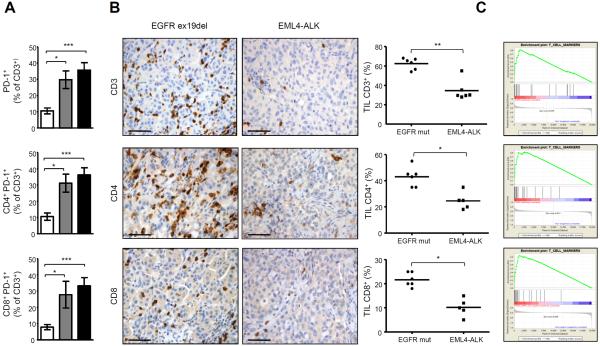
, n= 5 mice) and 16-week-old EML4-ALK mice (■, n=9 mice). B, Immunohistochemistry for CD3, CD4 and CD8 on a representative mutated EGFR (ex19del) patient (left panels) and a representative EML4-ALK positive NSCLC case (right panels). Scale bars, 100μm. Graphs show the percentages of CD3+, CD4+ and CD8+ cells in EML4-ALK positive NSCLC vs EGFR mutated patients. Horizontal bars represent means. C, Gene Set Enrichment Analysis (GSEA) for T cell markers based on gene expression profiling of human EML4-ALK NSCLC vs EGFR mutated NSCLC (L858R or EGFR-Del19) (FDR q-Value: 0.008, top panel) or vs K-RAS mutated NSCLC (FDR q-Value: 0.001, central panel) or vs KRAS/EGFR/ALK negative NSCLC (FDR q-Value: 0.00046, bottom panel). *, P<0.05; **, P<0.005; ***, P<0.0005.
Figure 5
Figure 5. Blockade of the PD-1/PD-L1 pathway restores the efficacy of the ALK vaccine against cells expressing high PD-L1
A, Western blot of H3122 cells treated with different crizotinib concentrations and collected at the indicated time points. Membranes were blotted with the indicated antibodies. B, PD-L1 protein expression was evaluated by flow cytometry in H3122 cells treated with 150nM crizotinib for 24 hours. C, PD-L1 mRNA expression was evaluated by qRT-PCR in crizotinib-treated cells. D, PD-L1 expression was evaluated by flow cytometry in ASB-XIV cells (Ctrl), EML4-ALK ASB-XIV (EML-ALK) and in EML4-ALK ASB-XIV transduced with PD-L1 (EML4-ALK/PD-L1). E, Mean tumor numbers in lungs from mice injected with the indicated ASB-XIV cells (n= 5 mice for each group). F, Mean tumor numbers in lungs from mice with the indicated treatments (n= 6–8 mice for each group). Data are represented as mean (±SEM). G and H, Quantification of volume changes compared to baseline tumors in ALK mice treated with control IgG (n=6 mice) or anti-PD-1 antibody (n=7 mice) at the end of treatment (G) and at 4 weeks after treatment suspension (H). Horizontal bars represent means. Data are from two independent experiments. *, P<0.05; **, P<0.005; ***, P<0.0005.
Figure 6
Figure 6. ALK vaccine is efficacious in combination with crizotinib treatment
A, Schematic representation of the ALK vaccination combined with crizotinib treatment in EML4-ALK mice. B, Cytotoxic activity in ALK vaccinated mice in combination with crizotinib. Horizontal bars represent means. C, Representative MRI of crizotinib-treated mice and crizotinib-treated plus vaccinated mice. Arrows indicate tumor recurrence in the same position. Arrowheads indicate new tumors. D and E, The number of tumors (D) and the tumor volume (E) were measured by MRI analysis at the indicated time points. Data are from two independent experiments. Data are represented as mean (±SEM). **, P<0.005; ***, P<0.0005.
Figure 7
Figure 7. ALK vaccine is effective in tumors with crizotinib resistant EML4-ALK mutants
A, Western Blot shows the expression of EML4-ALK wild-type or the EML4-ALK mutants (C1156Y, F1174L and L1196M) in ASB-XIV infected cells and in human ALK-rearranged NSCLC cell line (H3122). The lines between the blots indicate cut lanes on the same gel. B–E, Representative H&E sections of the lungs of control (Ctrl) and ALK vaccinated (Vax) mice at day 21 after injection i.v. of ASB-XIV cells infected with a GFP plasmid expressing EML4-ALK WT (B) or the EML4-ALK mutants C1156Y (C), L1196M (D) or F1174L (E). Histograms represent the number of tumors in control (Ctrl; n=3 mice for each EML4-ALK construct) and ALK vaccinated mice (Vax; n=3 mice for each EML4-ALK construct). Scale bars, 1mm. Data are from two independent experiments. Data are represented as mean (±SEM). *, P<0.05; **, P<0.005; ***, P<0.0005.

References

    1. Pao W, Girard N. New driver mutations in non-small-cell lung cancer. Lancet Oncol. 2011;12(2):175–80. - PubMed
    1. Berge EM, Doebele RC. Targeted therapies in non-small cell lung cancer: emerging oncogene targets following the success of epidermal growth factor receptor. Seminars in oncology. 2014;41(1):110–25. - PMC - PubMed
    1. Soda M, Choi YL, Enomoto M, Takada S, Yamashita Y, Ishikawa S, et al. Identification of the transforming EML4-ALK fusion gene in non-small-cell lung cancer. Nature. 2007;448(7153):561–6. - PubMed
    1. Chiarle R, Voena C, Ambrogio C, Piva R, Inghirami G. The anaplastic lymphoma kinase in the pathogenesis of cancer. Nature reviews Cancer. 2008;8(1):11–23. - PubMed
    1. Mano H. ALKoma: a cancer subtype with a shared target. Cancer discovery. 2012;2(6):495–502. - PubMed

Publication types

MeSH terms