. 2015 Sep 30:12:183.
doi: 10.1186/s12974-015-0404-7.

The expression levels of prolyl oligopeptidase responds not only to neuroinflammation but also to systemic inflammation upon liver failure in rat models and cirrhotic patients

Affiliations

The expression levels of prolyl oligopeptidase responds not only to neuroinflammation but also to systemic inflammation upon liver failure in rat models and cirrhotic patients

Jofre Tenorio-Laranga et al. J Neuroinflammation. .

Abstract

Background: Liver failure in experimental animals or in human cirrhosis elicits neuroinflammation. Prolyl oligopeptidase (PREP) has been implicated in neuroinflammatory events in neurodegenerative diseases: PREP protein levels are increased in brain glial cells upon neuroinflammatory insults, but the circulating PREP activity levels are decreased in multiple sclerosis patients in a process probably mediated by bioactive peptides. In this work, we studied the variation of PREP levels upon liver failure and correlated it with several inflammatory markers to conclude on the relation of PREP with systemic and/or neuroinflammation.

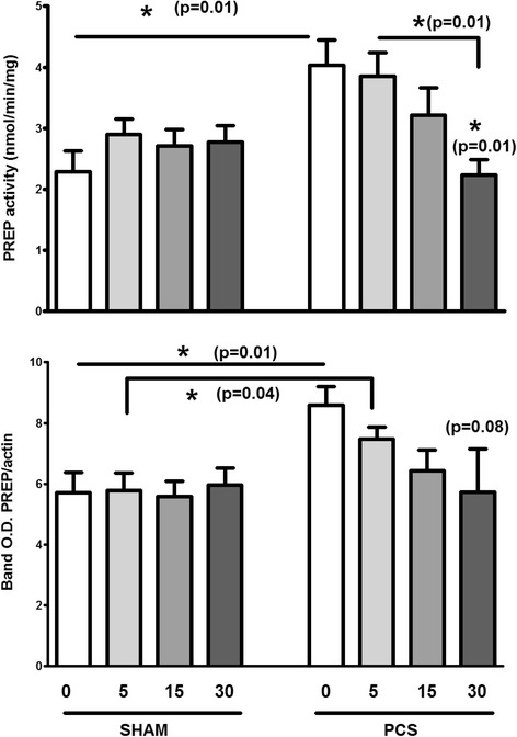
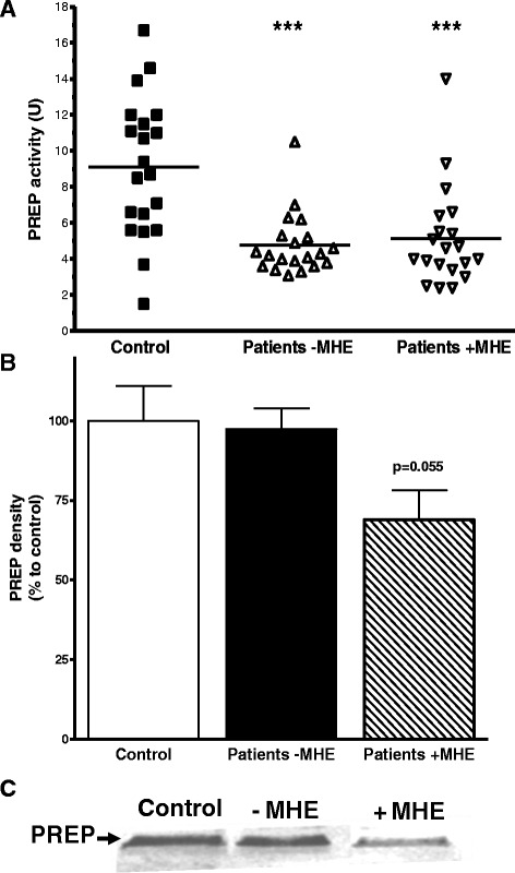
Methods: PREP enzymatic activity and protein levels measured with immunological techniques were determined in the brain and plasma of rats with portacaval shunt (PCS) and after treatment with ibuprofen. Those results were compared with the levels of PREP measured in plasma from cirrhotic patients with or without minimal hepatic encephalopathy (MHE). Levels of several pro-inflammatory cytokines and those of NO/cGMP homeostasis metabolites were measured in PCS rats and cirrhotic patients to conclude on the role of PREP in inflammation.

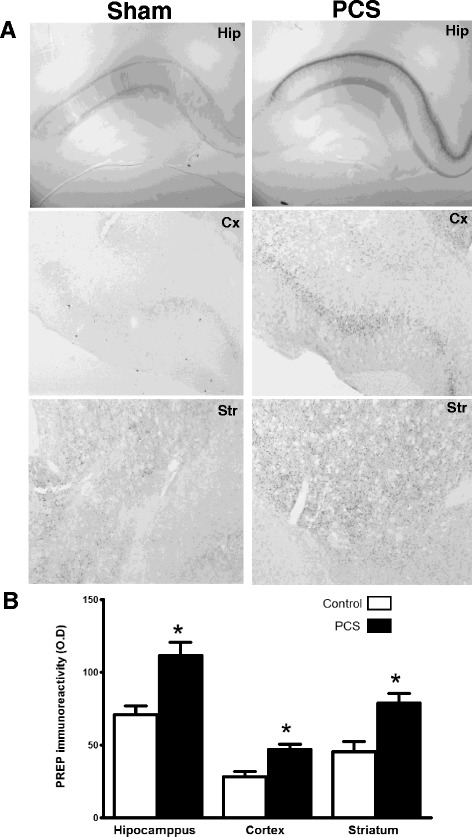
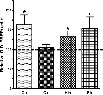
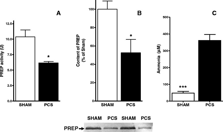
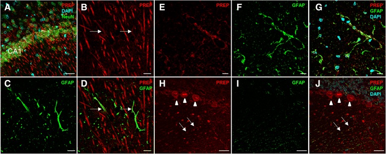
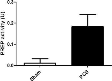
Results: In PCA rats, we found that PREP levels are significantly increased in the hippocampus, striatum and cerebellum, that in the cerebellum the PREP increase was significantly found in the extracellular space and that the levels were restored to those measured in control rats after administration of an anti-inflammatory agent, ibuprofen. In cirrhotic patients, circulatory PREP activity was found to correlate to systemic and neuroinflammatory markers and had a negative correlation with the severity of the disease, although no clear relation to MHE.

Conclusions: These results support the idea that PREP levels could be used as indicators of cirrhosis severity in humans, and using other markers, it might contribute to assessing the level of neuroinflammation in those patients. This work reports, for the first time, that PREP is secreted to the extracellular space in the cerebellum most probably due to glial activation and supports the role of the peptidase in the inflammatory response.


Figures

Fig. 1
Fig. 1
The expression of PREP in the brain of PCS rats is substantially increased as shown by a immunohistochemistry of sections of the hippocampus (Hip), frontal cortex (Cx) and striatum (Str), compared with sham operated rat brain. This increase in density was quantitated as represented in (b) (n = 4–5)
Fig. 2
Fig. 2
PREP expression was also detected increased by western blotting in the brain of PCS rats, compared with sham operated control rats (line at 100 %), especially in the cerebellum (Cb, n = 9), hippocampus (Hip, n = 6) and striatum (Str, n = 4). The increase was not significant in the cortex (Cx, n = 4)
Fig. 3
Fig. 3
PREP activity (a) and protein levels in plasma, determined by western blotting (b), are decreased (p < 0.05) in PCA rats (black bar, n = 4) compared with sham operated control rats (white bar, n = 5). Representative blots are show in the lower panel. c, ammonia levels in PCA are increased relative to control
Fig. 4
Fig. 4
PREP is expressed in neurons and astrocytes in hippocampus and cerebellum of PCS rats. PREP (red) is expressed mainly in the neurons of the CA1 region of the hippocampus visualized with NeuN (green) (a). High magnification images showed co-localization (arrows) of PREP (red) with astrocytes stained with glial fibrillary acidic protein (GFAP; green) in the hippocampus (bd), in the white matter of the cerebellum (eg) and the cerebellar cortex (hj). To note is the strong PREP expression in Purkinje cells (arrow heads in (h, j)) also co-localization with the glia Bergman (arrows). Nuclear marker: DAPI (cyan). Scale bar: bg 10 μm; a, hj 30 μm
Fig. 5
Fig. 5
Brain extracellular PREP activity measured in the dialysate of cerebellum microdialysis of sham operated control rats (white bar) and PCS rats (black bar) (n = 4)
Fig. 6
Fig. 6
PREP increased in PCA rats is recovered to control level after ibuprofen treatment. Groups of 4–5 animals of control sham operated rats (left) and PCA rats (left), were administrated with 0, 5, 15 and 30 mg/kg for 4 days, and striatal PREP levels of activity (upper graph) and protein (lower graph) were measured in both groups. The shade of grey in the bars increases with the increase on ibuprofen dose as showed. Statistical p values between selected bars are shown
Fig. 7
Fig. 7
a Average PREP activity in cirrhotic patients without (triangles) or with (inverted triangles) symptoms of MHE was significantly decreased compared with healthy subjects (***p < 0.001). b The levels of PREP protein in patients without MHE (black bar) showed no significant change, but these levels are apparently reduced in patients with MHE (hatch pattern bar), in comparison with PREP levels in control healthy subjects (white bar). A representative western blot is shown in c

References

    1. Frederick RT. Current concepts in the pathophysiology and management of hepatic encephalopathy. Gastroenterol Hepatol (N Y) 2011;7(4):222–33. - PMC - PubMed
    1. Das A, Dhiman RK, Saraswat VA, Verma M, Naik SR. Prevalence and natural history of subclinical hepatic encephalopathy in cirrhosis. J Gastroenterol Hepatol. 2001;16(5):531–5. doi: 10.1046/j.1440-1746.2001.02487.x. - DOI - PubMed
    1. Romero-Gómez M, Boza F, García-Valdecasas MS, García E, Aguilar-Reina J. Subclinical hepatic encephalopathy predicts the development of overt hepatic encephalopathy. Am J Gastroenterol. 2001;96(9):2718–23. doi: 10.1111/j.1572-0241.2001.04130.x. - DOI - PubMed
    1. Ferenci P, Lockwood A, Mullen K, Tarter R, Weissenborn K, Blei AT. Hepatic encephalopathy--definition, nomenclature, diagnosis, and quantification: final report of the working party at the 11th World Congresses of Gastroenterology, Vienna, 1998. Hepatology. 2002;35(3):716–21. doi: 10.1053/jhep.2002.31250. - DOI - PubMed
    1. Cauli O, Rodrigo R, Piedrafita B, Boix J, Felipo V. Inflammation and hepatic encephalopathy: ibuprofen restores learning ability in rats with portacaval shunts. Hepatology. 2007;46(2):514–9. doi: 10.1002/hep.21734. - DOI - PubMed

Publication types

MeSH terms

LinkOut - more resources