. 2015 Sep 29:6:186.
doi: 10.1186/s13287-015-0175-1.

Pre-differentiation of human neural stem cells into GABAergic neurons prior to transplant results in greater repopulation of the damaged brain and accelerates functional recovery after transient ischemic stroke

Affiliations

Pre-differentiation of human neural stem cells into GABAergic neurons prior to transplant results in greater repopulation of the damaged brain and accelerates functional recovery after transient ischemic stroke

Hima C S Abeysinghe et al. Stem Cell Res Ther. .

Abstract

Introduction: Despite attempts to prevent brain injury during the hyperacute phase of stroke, most sufferers end up with significant neuronal loss and functional deficits. The use of cell-based therapies to recover the injured brain offers new hope. In the current study, we employed human neural stem cells (hNSCs) isolated from subventricular zone (SVZ), and directed their differentiation into GABAergic neurons followed by transplantation to ischemic brain.

Methods: Pre-differentiated GABAergic neurons, undifferentiated SVZ-hNSCs or media alone were stereotaxically transplanted into the rat brain (n=7/group) 7 days after endothelin-1 induced stroke. Neurological outcome was assessed by neurological deficit scores and the cylinder test. Transplanted cell survival, cellular phenotype and maturation were assessed using immunohistochemistry and confocal microscopy.

Results: Behavioral assessments revealed accelerated improvements in motor function 7 days post-transplant in rats treated with pre-differentiated GABAergic cells in comparison to media alone and undifferentiated hNSC treated groups. Histopathology 28 days-post transplant indicated that pre-differentiated cells maintained their GABAergic neuronal phenotype, showed evidence of synaptogenesis and up-regulated expression of both GABA and calcium signaling proteins associated with neurotransmission. Rats treated with pre-differentiated cells also showed increased neurogenic activity within the SVZ at 28 days, suggesting an additional trophic role of these GABAergic cells. In contrast, undifferentiated SVZ-hNSCs predominantly differentiated into GFAP-positive astrocytes and appeared to be incorporated into the glial scar.

Conclusion: Our study is the first to show enhanced exogenous repopulation of a neuronal phenotype after stroke using techniques aimed at GABAergic cell induction prior to delivery that resulted in accelerated and improved functional recovery.


Figures
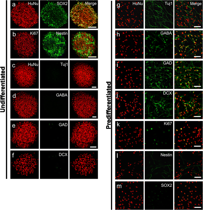
Fig. 1
Fig. 1
Undifferentiated hNSCs and predifferentiated cell phenotypes in vitro. Immunofluorescent confocal images of undifferentiated SVZ-hNSCs in culture expressed HuNu (red) and SOX2 (green) (a), and Ki67 (red) and Nestin (green) (b), with colocalization giving a yellow appearance. Undifferentiated SVZ-hNSCs immunopositive for HuNu (red) (cf) in culture were not observed to express Tuj1 (green) (c), GABA (green) (d), GAD (green) e, or DCX (green) f. Predifferentiated cells cultured for 7 days demonstrated immunoreactivity for HuNu (red) gm and colabeled with Tuj1 (green) (g), GABA (green) (h), GAD (green) (i), and DCX (green) (j). Predifferentiated cells HuNu (red) cultured for 7 days demonstrated little immunoreactivity for Ki67 (green) (k), Nestin (green) (l), or SOX2 (green) (m). Scale bar: (af) 100 μm, (gm) 50 μm. DCX doublecortin, GABA gamma-aminobutyric acid, GAD glutamate decarboxylase 65&67, HuNu human specific nuclear antigen, Tuj1 β-III tubulin
Fig. 2
Fig. 2
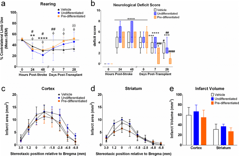
Functional outcome following transplantation. Effects of transplantation on contralateral limb use when rearing (a) in the cylinder test after ET-1-induced stroke. Data are mean ± standard error of the mean (SEM) expressed as a percentage of contralateral (impaired) forelimb use. Each rat acted as its own control; results following stroke were compared with 0-hour prestroke scores. ϕP <0.05, ϕϕP <0.01, ϕϕϕϕP <0.0001 relative to 0-hour poststroke baseline scores for vehicle-treated rats (n = 7); **P <0.01, ***P <0.001, ****P <0.0001 compared with 0-hour poststroke baseline scores for undifferentiated treated rats (n = 7); #P <0.05, ##P <0.01 relative to 0-hour poststroke baseline scores for predifferentiated treated rats (n = 7); δP <0.05, δδP <0.01 vehicle-treated rats compared with predifferentiated treated rats; φφP <0.01 vehicle-treated rats compared with undifferentiated treated rats; †P <0.05 predifferentiated treated rats compared with undifferentiated treated rats (two-way ANOVA followed by Bonferroni post test). Combined neurological deficit scores (b). Data presented as box plots include hinges extending from the 25th to 75th percentiles, the median line within the box and whiskers extending to the minimum and maximum values of the dataset (n = 7/group). *P <0.05, ***P <0.001, ****P <0.0001 relative to 0-hour poststroke baseline scores (n = 7/group); #P <0.05, ##P <0.01, ###P <0.001, ####P <0.0001 relative to pretransplant scores (Kruskal–Wallis ANOVA followed by Dunn’s test). Effect of transplanting vehicle, undifferentiated hNSCs, or predifferentiated cells on infarct area (c), (d) and total infarct volume (e) within the cortex and striatum. Data presented as mean ± SEM of infarct area measured at eight predetermined coronal planes through the brain (two-way ANOVA followed by Bonferroni post test)
Fig. 3
Fig. 3
Transplanted cells survive within the stroke affected brain. Total number (a) and percentage (b) of HuNu-positive cells remaining within cortical and striatal grafts from undifferentiated (n = 7) and predifferentiated (n = 7) treatment groups. Data presented as mean ± SEM. ****P <0.0001 relative to undifferentiated treated animals in the same region; ####P <0.0001 relative to undifferentiated grafts within the cortex; ϕP <0.05 relative to predifferentiated grafts within the striatum (two-way ANOVA followed by Bonferroni post test). Transplanted cells immunopositive for HuNu (red) (c, f) did not express apoptotic markers including Casp3 (green) (d), merged image (e), or TUNEL (g), with the level of TUNEL staining similar to the contralateral mirror image (h). Transplanted HuNu-positive (red) predifferentiated cells were associated with vWF-stained blood vessels (green) within infarcted brain regions (i). Many HuNu-positive undifferentiated hNSCs (red) were found within border regions consisting of GFAP-positive astrocytes (green) (j). Magnified immunofluorescent image (k) corresponds to box highlighted in (j) illustrating incorporation of undifferentiated hNSCs into the glial scar bordering the infarct. Scale bar: (ce) 20 μm, (fh) 100 μm, (i, k) 200 μm, (j) 100 μm. C contralateral hemisphere, Casp3 cleaved caspase-3, GFAP glial fibrillary acidic protein, HuNu human specific nuclear antigen, I ipsilateral hemisphere, TUNEL terminal transferase-mediated dUTP nick end-labeling, vWF von Willebrand factor
Fig. 4
Fig. 4
Undifferentiated hNSCs and predifferentiated cell phenotypes in vivo. Confocal immunofluorescent photomicrographs of undifferentiated hNSCs at 28 days post transplant within cortical border regions with orthogonal reconstructions. Undifferentiated hNSCs maintained expression of HuNu (red) and colabeled mainly with GFAP (a), Nestin (b), and Ki67 (c), with little expression of GABA (d) and Tuj1 (e) (green). Immunofluorescent confocal images of predifferentiated cells 28 days post transplant within the stroke-damaged cortex maintained expression of HuNu (red) colabeled with Tuj1 (f), GABA (g), Nestin (h), and Ki67 (i) (green), with lack of colocalization with GFAP (green) (j). Orthogonal reconstructions from confocal z-series are presented as viewed in xz (top) and yz (right) planes. HuNu immunoreactivity of undifferentiated hNSCs was observed completely surrounded by GFAP and Nestin, while HuNu immunoreactivity of predifferentiated cells was observed completely surrounded by Tuj1 and GABA. Scale bar: (ae) 50 μm, orthogonal images 5 μm; (fj) 20 μm, orthogonal images 5 μm. GABA gamma-aminobutyric acid, GFAP glial fibrillary acidic protein, HuNu human specific nuclear antigen, Tuj1 β-III tubulin
Fig. 5
Fig. 5
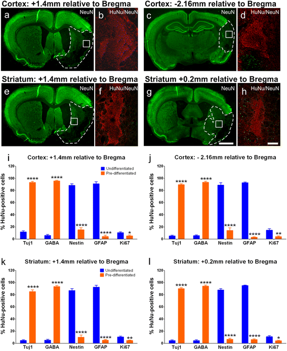
Phenotypic profile of transplanted undifferentiated hNSCs versus predifferentiated cells. Coronal sections immunostained with NeuN (green) highlight undamaged brain regions with lack of staining within stroke affected areas (white dotted line) at cortical graft sites +1.4 mm and –2.16 mm relative to the bregma (a, c) and striatal graft sites +1.4 mm and +0.2 mm relative to the bregma (e, g); white boxes depict graft location. Representative images of graft sites stained with HuNu (red) and NeuN (green) (b, d, f, h) from regions highlighted by white boxes (a, c, e, g). Numbers of HuNu-positive cells coexpressing Tuj1, GABA, Nestin, GFAP, and Ki67 from undifferentiated hNSC-treated (n = 7) and predifferentiated cell-treated (n = 7) animals from cortical graft sites; +1.4 mm (i) and –2.16 mm (j) relative to the bregma; and striatal graft sites +1.4 mm (k) and +0.2 mm (l) relative to the bregma. Numbers of cells are presented as a percentage of the total number of HuNu-positive cells counted. Data are mean ± SEM. ***P <0.001, ****P <0.0001 compared with undifferentiated hNSC counts (two-way ANOVA with Bonferroni post test). Scale bar: (a, c, e, g) 2000 μm, (b, d, f, h) 200 μm. GABA gamma-aminobutyric acid, GFAP glial fibrillary acidic protein, HuNu human specific nuclear antigen, NeuN neuron specific nuclear antigen, Tuj1 β-III tubulin
Fig. 6
Fig. 6
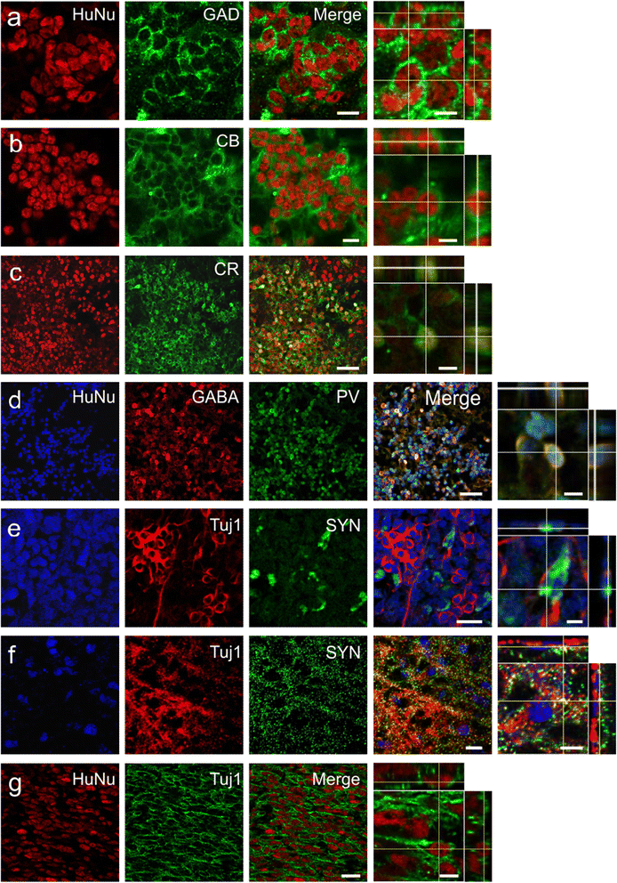
Further maturation of predifferentiated cells 28 days post transplant. Confocal photomicrographs of predifferentiated cells 28 days post transplant within the stroke-damaged brain expressed HuNu (red) double labeled with either GAD65/67 (green) (a), calbindin-D28k (CB; green) (b), or calretinin (CR, green) (c). Predifferentiated cells expressing HuNu (blue) triple-labeled with parvalbumin (PV, green) and GABA (red) (d), or triple-labeled with Tuj1 (red) and presynaptic vesicle protein synaptophysin (SYN, green) within the cortical core (e) and border (f) regions. Some predifferentiated cells expressing HuNu (red) grafted to border regions appeared to extend long neurites expressing Tuj1 (green) (g). Orthogonal reconstructions from confocal z-series are presented as viewed in xz (top) and yz (right) planes. Scale bar: (a, b) 10 μm, orthogonal image 5 μm; (c, d) 40 μm, orthogonal image 5 μm; (e, f) 10 μm, orthogonal image 5 μm; (g) 20 μm, orthogonal image 5 μm. GABA gamma-aminobutyric acid, GAD glutamate decarboxylase 65&67, HuNu human specific nuclear antigen, Tuj1 β-III tubulin
Fig. 7
Fig. 7
Predifferentiated cells grafted to the core infarct in vivo. Predifferentiated cell graft (HuNu, red) (a) located within the infarcted core region was surrounded by GFAP-positive astrocytes on one side (green) (b) that form the glial scar border (arrow) and merge (c), with dispersion of HuNu-positive cells observed in the core furthest away from the scar (arrowheads). Negative control; omission of primary antibodies (df). Scale bar: (af) 100 μm. GFAP glial fibrillary acidic protein, HuNu human specific nuclear antigen
Fig. 8
Fig. 8
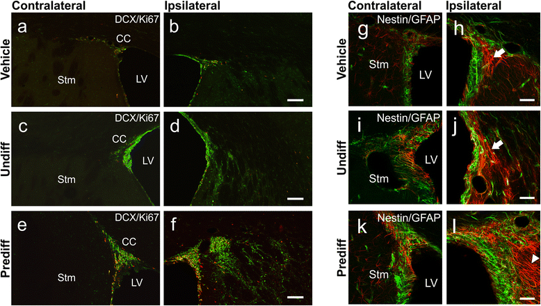
Effect of treatment on endogenous neurogenesis and radial glial populations within the SVZ. Immunofluorescent images of proliferating cells (Ki67; red) and migrating immature neuroblasts (DCX; green) within the contralateral and ipsilateral SVZ of vehicle-treated (a, b), undifferentiated hNSC-treated (c, d), and predifferentiated cell-treated (e, f) animals. Nestin (green; arrowheads) and GFAP (red) immunopositive cells (coexpression giving a yellow appearance; arrows) within the contralateral and ipsilateral SVZ of vehicle-treated (g, h), undifferentiated treated (i, j), and predifferentiated treated (k, l) animals. All images were taken at the same anatomical location from animals with similar infarct volumes. Scale bar: (af) 500 μm, (gl) 50 μm. CC corpus callosum, DCX doublecortin, GFAP glial fibrillary acidic protein, LV lateral ventricle, Prediff predifferentiated treated, Stm Striatum, Undiff undifferentiated hNSC treated

Similar articles

Cited by

References

    1. Thom T, Haase N, Rosamond W, Howard VJ, Rumsfeld J, et al. Heart disease and stroke statistics—2006 update: a report from the American Heart Association Statistics Committee and Stroke Statistics Subcommittee. Circulation. 2006;113:e85–e151. doi: 10.1161/CIRCULATIONAHA.105.171600. - DOI - PubMed
    1. Molina CA. Reperfusion therapies for acute ischemic stroke: current pharmacological and mechanical approaches. Stroke. 2011;42:S16–S19. doi: 10.1161/STROKEAHA.110.598763. - DOI - PubMed
    1. Zhang RL, Zhang ZG, Chopp M. Ischemic stroke and neurogenesis in the subventricular zone. Neuropharmacology. 2008;55:345–352. doi: 10.1016/j.neuropharm.2008.05.027. - DOI - PMC - PubMed
    1. Dancause N, Barbay S, Frost SB, Plautz EJ, Chen D, et al. Extensive cortical rewiring after brain injury. J Neurosci. 2005;25:10167–10179. doi: 10.1523/JNEUROSCI.3256-05.2005. - DOI - PMC - PubMed
    1. Nelles G. Cortical reorganization-effects of intensive therapy. Restor Neurol Neurosci. 2004;22:239–244. - PubMed

Publication types

MeSH terms