. 2015 Dec 4;290(49):29127-39.
doi: 10.1074/jbc.M115.689000. Epub 2015 Sep 29.

Structural Basis for Ligand Recognition and Functional Selectivity at Angiotensin Receptor

Affiliations

Structural Basis for Ligand Recognition and Functional Selectivity at Angiotensin Receptor

Haitao Zhang et al. J Biol Chem. .

Abstract

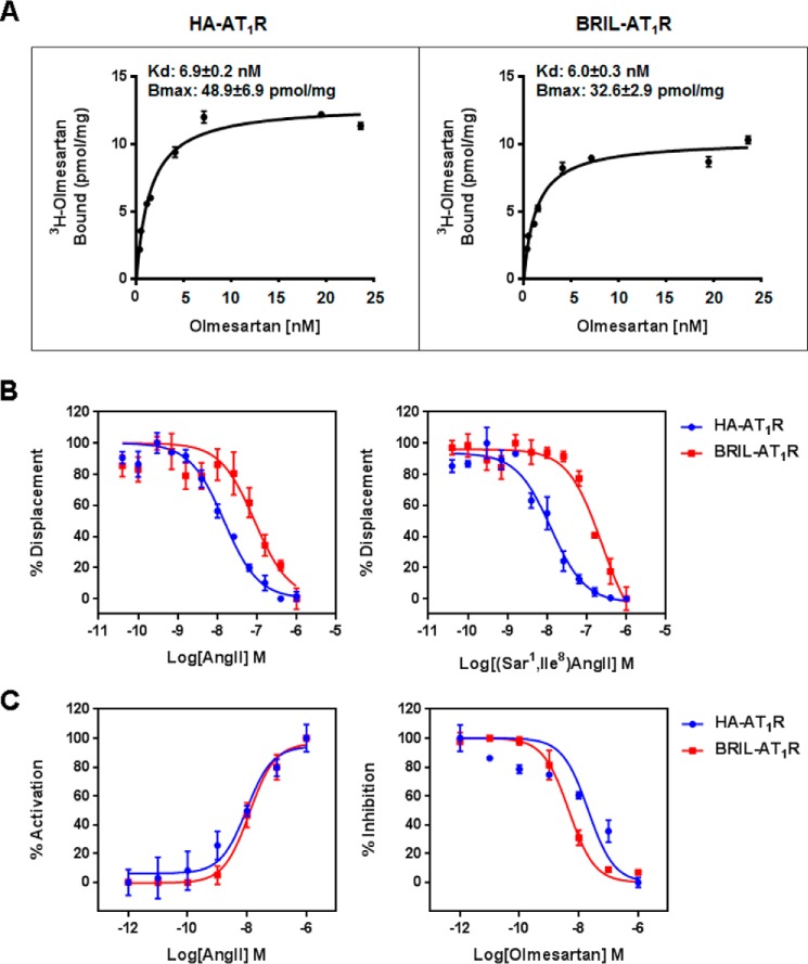
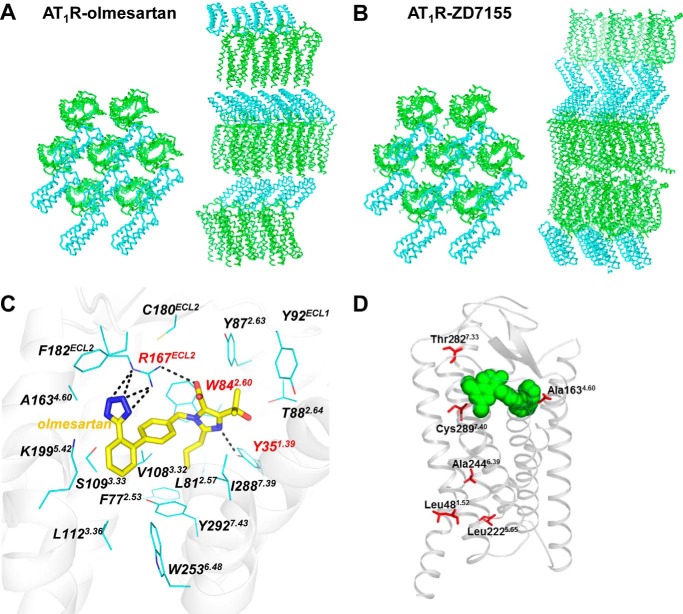
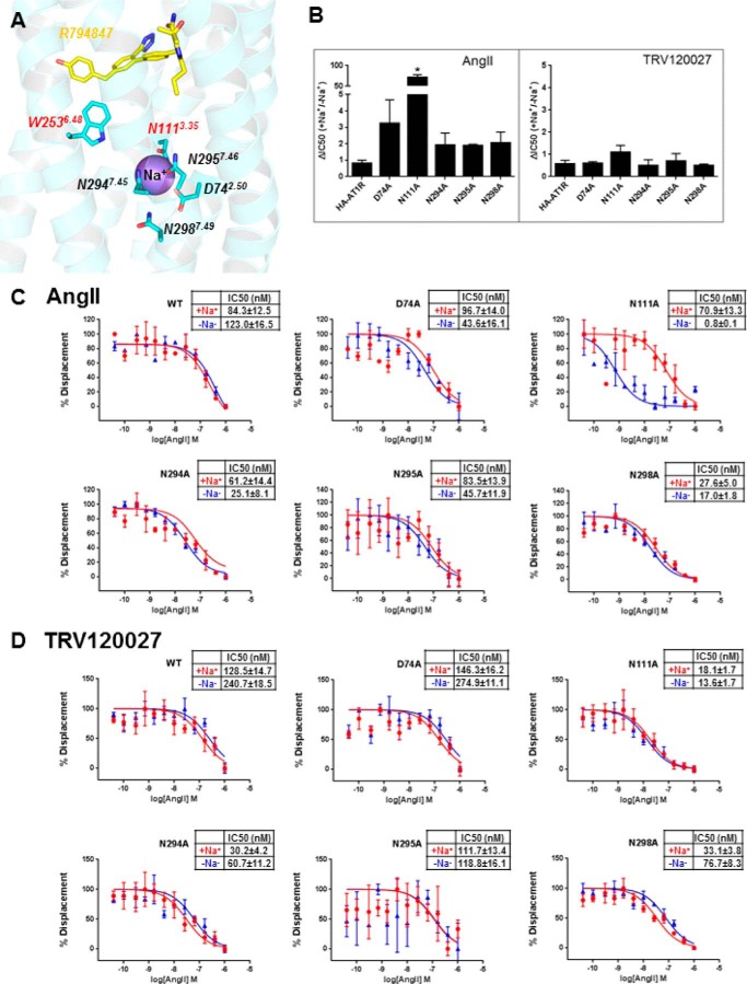
Angiotensin II type 1 receptor (AT1R) is the primary blood pressure regulator. AT1R blockers (ARBs) have been widely used in clinical settings as anti-hypertensive drugs and share a similar chemical scaffold, although even minor variations can lead to distinct therapeutic efficacies toward cardiovascular etiologies. The structural basis for AT1R modulation by different peptide and non-peptide ligands has remained elusive. Here, we report the crystal structure of the human AT1R in complex with an inverse agonist olmesartan (Benicar(TM)), a highly potent anti-hypertensive drug. Olmesartan is anchored to the receptor primarily by the residues Tyr-35(1.39), Trp-84(2.60), and Arg-167(ECL2), similar to the antagonist ZD7155, corroborating a common binding mode of different ARBs. Using docking simulations and site-directed mutagenesis, we identified specific interactions between AT1R and different ARBs, including olmesartan derivatives with inverse agonist, neutral antagonist, or agonist activities. We further observed that the mutation N111(3.35)A in the putative sodium-binding site affects binding of the endogenous peptide agonist angiotensin II but not the β-arrestin-biased peptide TRV120027.

Keywords: G protein-coupled receptor (GPCR); angiotensin; biased ligands; crystal structure; hypertension; protein-drug interaction; sodium ion.


Figures

FIGURE 1.
FIGURE 1.
Comparisons of the ligand binding and functional activities between the wild-type (HA-AT1R) and crystallized construct (BRIL-AT1R). A, binding of olmesartan to the wild-type HA-AT1R and crystallized BRIL-AT1R. Binding studies were performed using isolated membranes from transiently transfected COS1 cells. Saturation binding curves were measured using [3H]olmesartan, and the corresponding Kd and Bmax values were obtained by non-linear curve fitting. B, displacement of [3H]olmesartan with the agonist AngII and the antagonist [Sar1,Ile8]AngII in the wild-type HA-AT1R and the crystallized BRIL-AT1R. Binding studies were performed using isolated membranes from transiently transfected COS1 cells. Competition binding curves for peptide agonist AngII and peptide antagonist [Sar1,Ile8]AngII were generated, and the corresponding IC50 values were calculated. The IC50 values for AngII to inhibit olmesartan binding were 1.8 ± 0.2 and 18.4 ± 2.3 nm for HA-AT1R and BRIL-AT1R, respectively. The IC50 values for [Sar1,Ile8]AngII to inhibit olmesartan binding were 1.2 ± 0.1 and 25.5 ± 4.9 nm for HA-AT1R and BRIL-AT1R, respectively. C, intracellular calcium responses of wild-type HA-AT1R and BRIL-AT1R. AngII and olmesartan dose-response curves for HA-AT1R and BRIL-AT1R are shown. For the antagonist dose response, cells were treated with 0–10 μm concentrations of olmesartan followed by stimulation with 100 nm AngII. The EC50 values for AngII dose response were 10.2 ± 3.2 and 11.9 ± 3.1 nm for HA-AT1R and BRIL-AT1R, respectively. The IC50 values for olmesartan to inhibit AngII response were between 23.9 ± 8.0 and 4.4 ± 0.4 nm for HA-AT1R and BRIL-AT1R, respectively. All results above are presented as mean ± S.E. and represent three experiments performed in triplicate.
FIGURE 2.
FIGURE 2.
Crystal structure of AT1R-olmesartan. A and B, crystal packing comparison of the two AT1R structures bound to the inverse agonist olmesartan (this work) and the antagonist ZD7155 (Protein Data Bank code 4YAY). Crystal packing views perpendicularly (left) and parallel (right) to the membrane are shown for the two N-terminal BRIL-fused AT1R structures bound to different ligands, with AT1R shown as green ribbons and BRIL shown as cyan ribbons. C, interactions between AT1R and olmesartan were determined by crystal structure. D, three SNP variations, A1634.60T, T2827.33M, and C2897.40W, located in close proximity to the AT1R orthosteric binding pocket may differentially affect efficacies of specific ARBs in individuals carrying these SNPs via changes in direct or indirect interactions with the pocket.
FIGURE 3.
FIGURE 3.
Olmesartan derivatives with different interactions and functions toward AT1R. A, IP signaling assays identified that olmesartan and R781253 are inverse agonists; R239470 is a neutral antagonist, and R794847 is a weak partial agonist of the human AT1R. Statistical analysis was performed by unpaired two-tailed t test, in which p values less than 0.05 were considered statistically significant. Olmesartan, p = 0.037; R781253, p = 0.042; R794847, p = 0.048, and AngII, p = 0.024. Results are presented as means ± S.E. and represent three experiments performed in triplicate. B, docking simulations showed conserved binding modes of olmesartan (cyan), R239470 (blue), R781253 (green), and R794847 (red) in the AT1R ligand-binding pocket. C, schematic representations for interactions of olmesartan and its derivatives with AT1R are shown with hydrogen bonds/salt bridges as red dashed lines. B and C, three critical residues for binding of all four ligands are highlighted in red; the residues discriminating ligand binding are labeled in green (for R239470 and R794847) and cyan (for R781253 and R794847).
FIGURE 4.
FIGURE 4.
Allosteric modulation of AngII and TRV120027 binding by sodium ion in sodium-binding pocket mutants. A, structure of AT1R with docked R794847 ligand shows that the toggle switch Trp-2536.48 belongs to the sodium-binding pocket and interacts with the 4-hydroxybenzyl pharmacophore of R794847. B–D, competition binding assays were performed under equilibrium conditions, with 2 nm [3H]olmesartan and concentrations of AngII or TRV120027 ranging between 0.04 and 1000 nm. The binding buffer used in the sodium containing (+Na+) experiments (red lines) was 140 mm NaCl, 5.4 mm KCl, 1 mm EDTA, 0.006% BSA, 25 mm HEPES, pH 7.4. The binding buffer used in the sodium deficient (−Na+) experiments (blue lines) was 140 mm N-methyl d-glucamine, 5.4 mm KCl, 1 mm EDTA, 0.006% BSA, 25 mm HEPES, pH 7.4. Statistical analysis was performed by unpaired two-tailed t test, in which p values less than 0.05 (asterisks) were considered statistically significant. N111A, p = 0.004. Results are presented as mean ± S.E. and represent three experiments performed in triplicate.

References

    1. Zaman M. A., Oparil S., and Calhoun D. A. (2002) Drugs targeting the renin-angiotensin-aldosterone system. Nat. Rev. Drug Discov. 1, 621–636 - PubMed
    1. Balakumar P., and Jagadeesh G. (2014) Structural determinants for binding, activation, and functional selectivity of the angiotensin AT1 receptor. J. Mol. Endocrinol. 53, R71–R92 - PubMed
    1. de Gasparo M., Catt K. J., Inagami T., Wright J. W., and Unger T. (2000) International union of pharmacology. XXIII. The angiotensin II receptors. Pharmacol. Rev. 52, 415–472 - PubMed
    1. Oliveira L., Costa-Neto C. M., Nakaie C. R., Schreier S., Shimuta S. I., and Paiva A. C. (2007) The angiotensin II AT1 receptor structure-activity correlations in the light of rhodopsin structure. Physiol. Rev. 87, 565–592 - PubMed
    1. Burnier M., and Brunner H. R. (2000) Angiotensin II receptor antagonists. Lancet 355, 637–645 - PubMed

Publication types

MeSH terms

Associated data